Era of Bloodless Surgery: Spotlights on Hemostasic Materials and Techniques
- Affiliations
-
- 1Department of Surgery, Hanyang University College of Medicine, Seoul, Korea.
- 2Department of Obstetrics and Gynecology, Ilsan Paik Hospital, Inje University, Goyang, Korea. j1985@paik.ac.kr
- KMID: 2407797
- DOI: http://doi.org/10.7599/hmr.2018.38.1.3
Abstract
- Ever since mankind has had blood, efforts to stop bleeding have never ceased and so numerous methods for hemostasis have been developed. In recent decades, minimally invasive surgical techniques have led patients to less-bleeding surgery but, hemostatic agents, devices and techniques still play an important role in medical side. A number of hemostatic agents and devices have been developed and they can be classified by their mechanism of action. That classification of the coagulants includes mechanisms with physical, caustic, bio-physical, biologic actions. Hemostatic devices are divided into categories such as dressings, glue, clips, electrocoagulations and so on. Based on the concept of minimally invasive surgical procedures, variously developed surgical techniques are divided by the number of ports used and auxiliary instruments. However, there are advantages and disadvantages to each of the hemostatic agents and minimally invasive methods, and the belief in the classical method also prevents the application of new hemostatic methods. The knowledge and understanding of the benefits and costs of these newly developed hemostatic methods will make it easier for medical personnel to manage patient's blood.
MeSH Terms
Figure
-

Fig. 1 (A) Arteriolar vasoconstriction occurs immediately by the reflex mechanism of the nervous system right after vascular injury, which can be enhanced by endothelin, a potent vasoconstrictor released from the endothelial cells constituting the vessel wall. (B) Platelets bind to the von Willebrand factor and attach to the extracellular matrix at the site of injury, after that, they change their appearance and promote further recruitment and aggregation of platelets by releasing granules such as ADP and Tx A2. (C) Tissue factor released from vascular endothelial cells expresses the platelet phospholipid complex. Through the coagulation cascade, they eventually activate thrombin and ultimately make the fibrin polymer to form thrombus. (D) During this period, the platelet plug contains trapped neutrophils and RBCs in the blood vessels, showing permanent plugs and preventing further bleeding. In the absence of vascular injury or complete thrombus formation, the endothelial cells secrete t-PA and thrombomodulin, which inhibit platelet adhesion and aggregation, to exert antithrombotic effects that lead limitation of hemostasis.
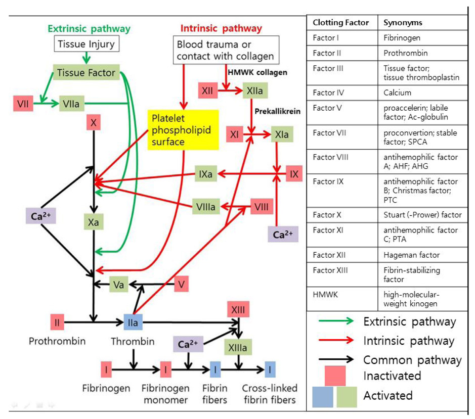
Fig. 2 The main role of extrinsic pathway that follows green arrows is generation of thrombin burst and thrombin burst is processed by thrombin, the most important coagulation factor of the coagulation cascade. It starts with tissue factors from damaged tissue and coagulation factor VII that moves around in blood plasma in a higher concentration than any other factors. The intrinsic pathway of red lines is activated by the primary complex formation on collagen with prekallikrein, high-molecular-weight kininogen (HMWK) and coagulation factor XII, known as Hageman factor. This intrinsic pathway seems to have association with inflammation and innate immunity. These two courses of coagulation pathway are the intrinsic pathway and the extrinsic pathway which both are concluded in the same common pathway along the black line to form cross-linked fibrin fibers from fibrinogen. SPCA, Serum prothrombin conversion accelerator; AHF, antihemophilic factor; AHG, antihemophilic globulin; PTC, plasma thromboplastin component; PTA, plasma thromboplastin antecedent.
Cited by 1 articles
-
Cutting-Edge Technologies for Patient Blood Management
Dongho Choi
Hanyang Med Rev. 2018;38(1):1-2. doi: 10.7599/hmr.2018.38.1.1.
Reference
-
1. Robbins SL, Kumar V, Cotran RS. Robbins and Cotran pathologic basis of disease. 8th ed. Philadelphia, PA: Saunders/Elsevier;2010.2. Hall JE, Guyton AC. Guyton and Hall textbook of medical physiology. 12th ed. Philadelphia, Pa.: Saunders/Elsevier;2011.3. Marieb EN, Hoehn K. Human anatomy & physiology. 8th ed. San Francisco: Benjamin Cummings;2010.4. Boon GD. An overview of hemostasis. Toxicol Pathol. 1993; 21:170–179.
Article5. Porrett PM, Atluri P, Karakousis GC, Roses RE, Drebin JA. The surgical review: an integrated basic and clinical science study guide. 4th ed.6. Zdanowicz MM. Essentials of pathophysiology for pharmacy. Boca Raton: CRC Press;2003.7. Li Z, Delaney MK, O'Brien KA, Du X. Signaling during platelet adhesion and activation. Arterioscler Thromb Vasc Biol. 2010; 30:2341–2349.
Article8. Long AT, Kenne E, Jung R, Fuchs TA, Renne T. Contact system revisited: an interface between inflammation, coagulation, and innate immunity. J Thromb Haemost. 2016; 14:427–437.
Article9. Bahn SL, Mursch PI. The effects of cold on hemostasis. Oral Surg Oral Med Oral Pathol. 1980; 49:294–300.
Article10. Larson PO. Topical hemostatic agents for dermatologic surgery. J Dermatol Surg Oncol. 1988; 14:623–632.11. Tan TC, Black PM. Sir Victor Horsley (1857-1916): pioneer of neurological surgery. Neurosurgery. 2002; 50:607–611. discussion 11-2.
Article12. Savva A, Taylor MJ, Beatty CW. Management of cerebrospinal fluid leaks involving the temporal bone: report on 92 patients. Laryngoscope. 2003; 113:50–56.
Article13. Schonauer C, Tessitore E, Barbagallo G, Albanese V, Moraci A. The use of local agents: bone wax, gelatin, collagen, oxidized cellulose. Eur Spine J. 2004; 13:Suppl 1. S89–S96.
Article14. Wang MY, Armstrong JK, Fisher TC, Meiselman HJ, McComb GJ, Levy ML. A new, pluronic-based, bone hemostatic agent that does not impair osteogenesis. Neurosurgery. 2001; 49:962–967. discussion 8.
Article15. Wilkinson HA, Baker S, Rosenfeld S. Gelfoam paste in experimental laminectomy and cranial trephination: hemostasis and bone healing. J Neurosurg. 1981; 54:664–667.16. Landry JR, Kanat IO. Considerations in topical hemostasis. J Am Podiatr Med Assoc. 1985; 75:581–585.
Article17. Wellisz T, An YH, Wen X, Kang Q, Hill CM, Armstrong JK. Infection rates and healing using bone wax and a soluble polymer material. Clin Orthop Relat Res. 2008; 466:481–486.
Article18. Frantz VK. Absorbable Cotton, Paper and Gauze: (Oxidized Cellulose). Ann Surg. 1943; 118:116–126.19. Fukuyama Y, Kawarai S, Tezuka T, Kawabata A, Maruo T. The palliative efficacy of modified Mohs paste for controlling canine and feline malignant skin wounds. Vet Q. 2016; 36:176–182.
Article20. Kakimoto M, Tokita H, Okamura T, Yoshino K. A chemical hemostatic technique for bleeding from malignant wounds. J Palliat Med. 2010; 13:11–13.
Article21. Yanazume S, Douzono H, Yanazume Y, Iio K, Douchi T. New hemostatic method using Mohs' paste for fatal genital bleeding in advanced cervical cancer. Gynecol Oncol Case Rep. 2013; 4:47–49.
Article22. Scher KS, Coil JA, Jr . Effects of oxidized cellulose and microfibrillar collagen on infection. Surgery. 1982; 91:301–304.23. Wagner WR, Pachence JM, Ristich J, Johnson PC. Comparative in vitro analysis of topical hemostatic agents. J Surg Res. 1996; 66:100–108.24. Abbott WM, Austen WG. The effectiveness and mechanism of collagen-induced topical hemostasis. Surgery. 1975; 78:723–729.25. Alexander JM, Rabinowitz JL. Microfibrillar collagen (Avitene) as a hemostatic agent in experimental oral wounds. J Oral Surg. 1978; 36:202–205.26. Uluyol S. Effects of silver nitrate cauterization on middle turbinate synechia after endoscopic sinus surgery. Otolaryngol Head Neck Surg. 2017; 157:515–518.
Article27. Montgomery BD, Boorjian SA, Ziegelmann MJ, Joyce DD, Linder BJ. Intravesical silver nitrate for refractory hemorrhagic cystitis. Turk J Urol. 2016; 42:197–201.
Article28. Palm MD, Altman JS. Topical hemostatic agents: a review. Dermatol Surg. 2008; 34:431–445.
Article29. Parker RK, Dinehart SM. Hints for hemostasis. Dermatol Clin. 1994; 12:601–606.
Article30. Naftalin LW, Yagiela JA. Vasoconstrictors: indications and precautions. Dent Clin North Am. 2002; 46:733–746. ix
Article31. Spalding A, Vaitkevicius H, Dill S, MacKenzie S, Schmaier A, Lockette W. Mechanism of epinephrine-induced platelet aggregation. Hypertension. 1998; 31:603–607.
Article32. Anschuetz L, Bonali M, Guarino P, Fabbri FB, Alicandri-Ciufelli M, Villari D, et al. Management of bleeding in exclusive endoscopic ear surgery: pilot clinical experience. Otolaryngol Head Neck Surg. 2017; 700–706.
Article33. Rousou J, Levitsky S, Gonzalez-Lavin L, Cosgrove D, Magilligan D, Weldon C, et al. Randomized clinical trial of fibrin sealant in patients undergoing resternotomy or reoperation after cardiac operations. A multicenter study. J Thorac Cardiovasc Surg. 1989; 97:194–203.
Article34. Spotnitz WD. Fibrin Sealant: the only approved hemostat, sealant, and adhesive-a laboratory and clinical perspective. ISRN Surg. 2014; 2014:203943.
Article35. Martinowitz U, Schulman S. Fibrin sealant in surgery of patients with a hemorrhagic diathesis. Thromb Haemost. 1995; 74:486–492.
Article36. Currie LJ, Sharpe JR, Martin R. The use of fibrin glue in skin grafts and tissue-engineered skin replacements: a review. Plast Reconstr Surg. 2001; 108:1713–1726.
Article37. Morikawa T. Tissue sealing. Am J Surg. 2001; 182:29S–35S.
Article38. Thompson DF, Letassy NA, Thompson GD. Fibrin glue: a review of its preparation, efficacy, and adverse effects as a topical hemostat. Drug Intell Clin Pharm. 1988; 22:946–952.
Article39. Bhanot S, Alex JC. Current applications of platelet gels in facial plastic surgery. Facial Plast Surg. 2002; 18:27–33.
Article40. Pomerantz J, Dutton JM. Platelet gel for endoscopic sinus surgery. Ann Otol Rhinol Laryngol. 2005; 114:699–704.
Article41. Achneck HE, Sileshi B, Jamiolkowski RM, Albala DM, Shapiro ML, Lawson JH. A comprehensive review of topical hemostatic agents: efficacy and recommendations for use. Ann Surg. 2010; 251:217–228.42. Pusateri AE, Kheirabadi BS, Delgado AV, Doyle JW, Kanellos J, Uscilowicz JM, et al. Structural design of the dry fibrin sealant dressing and its impact on the hemostatic efficacy of the product. J Biomed Mater Res B Appl Biomater. 2004; 70:114–121.
Article43. Neuffer MC, McDivitt J, Rose D, King K, Cloonan CC, Vayer JS. Hemostatic dressings for the first responder: a review. Mil Med. 2004; 169:716–720.
Article44. Alam HB, Burris D, DaCorta JA, Rhee P. Hemorrhage control in the battlefield: role of new hemostatic agents. Mil Med. 2005; 170:63–69.
Article45. Kulling D, Vournakis JN, Woo S, Demcheva MV, Tagge DU, Rios G, et al. Endoscopic injection of bleeding esophageal varices with a poly-N-acetyl glucosamine gel formulation in the canine portal hypertension model. Gastrointest Endosc. 1999; 49:764–771.
Article46. Rabea EI, Badawy ME, Stevens CV, Smagghe G, Steurbaut W. Chitosan as antimicrobial agent: applications and mode of action. Biomacromolecules. 2003; 4:1457–1465.
Article47. Noh JH, Lee JW, Nam YJ, Choi KY. Is intraoperative use of quikclot combat gauze effective for hemostasis after total knee arthroplasty? Clin Orthop Surg. 2017; 9:43–49.
Article48. Rhee P, Brown C, Martin M, Salim A, Plurad D, Green D, et al. QuikClot use in trauma for hemorrhage control: case series of 103 documented uses. J Trauma. 2008; 64:1093–1099.
Article49. Eaglstein WH, Sullivan T. Cyanoacrylates for skin closure. Dermatol Clin. 2005; 23:193–198.
Article50. Lo GH, Lai KH, Cheng JS, Chen MH, Chiang HT. A prospective, randomized trial of butyl cyanoacrylate injection versus band ligation in the management of bleeding gastric varices. Hepatology. 2001; 33:1060–1064.
Article51. Spotnitz WD, Burks S. Hemostats, sealants, and adhesives III: a new update as well as cost and regulatory considerations for components of the surgical toolbox. Transfusion. 2012; 52:2243–2255.
Article52. Torchiana DF. Polyethylene glycol based synthetic sealants: potential uses in cardiac surgery. J Card Surg. 2003; 18:504–506.53. Saunders MM, Baxter ZC, Abou-Elella A, Kunselman AR, Trussell JC. BioGlue and Dermabond save time, leak less, and are not mechanically inferior to two-layer and modified one-layer vasovasostomy. Fertil Steril. 2009; 91:560–565.
Article54. Biggs G, Hafron J, Feliciano J, Hoenig DM. Treatment of splenic injury during laparoscopic nephrectomy with BioGlue, a surgical adhesive. Urology. 2005; 66:882.
Article55. Luk A, David TE, Butany J. Complications of Bioglue postsurgery for aortic dissections and aortic valve replacement. J Clin Pathol. 2012; 65:1008–1012.
Article56. Passage J, Jalali H, Tam RK, Harrocks S, O'Brien MF. BioGlue Surgical Adhesive--an appraisal of its indications in cardiac surgery. Ann Thorac Surg. 2002; 74:432–437.
Article57. Fujita J, Takiguchi S, Nishikawa K, Kimura Y, Imamura H, Tamura S, et al. Randomized controlled trial of the LigaSure vessel sealing system versus conventional open gastrectomy for gastric cancer. Surg Today. 2014; 44:1723–1729.
Article58. Santini M, Fiorelli A, Messina G, Mazzella A, Accardo M. The feasibility of ligasure to create intestinal anastomosis: results of ex vivo study. Surg Innov. 2015; 22:266–273.
Article59. Diamantis T, Kontos M, Arvelakis A, Syroukis S, Koronarchis D, Papalois A, et al. Comparison of monopolar electrocoagulation, bipolar electrocoagulation, Ultracision, and Ligasure. Surg Today. 2006; 36:908–913.
Article60. Solaini L, Arru L, Merigo G, Tomasoni M, Gheza F, Tiberio GA. Advanced sealing and dissecting devices in laparoscopic adrenal surgery. Jsls. 2013; 17:622–626.
Article61. Prgomet D, Janjanin S, Bilic M, Prstacic R, Kovac L, Rudes M, et al. A prospective observational study of 363 cases operated with three different harmonic scalpels. Eur Arch Otorhinolaryngol. 2009; 266:1965–1970.
Article62. Huang J, Yu Y, Wei C, Qin Q, Mo Q, Yang W. Harmonic scalpel versus electrocautery dissection in modified radical mastectomy for breast cancer: a meta-analysis. PLoS One. 2015; 10:142–271.
Article63. Cheng H, Hsiao CW, Clymer JW, Schwiers ML, Tibensky BN, Patel L, et al. Gastrectomy and D2 lymphadenectomy for gastric cancer: a meta-analysis comparing the harmonic scalpel to conventional techniques. Int J Surg Oncol. 2015; 2015:397260.
Article64. Allaix ME, Arezzo A, Giraudo G, Arolfo S, Mistrangelo M, Morino M. The thunderbeat and other energy devices in laparoscopic colorectal resections: analysis of outcomes and costs. J Laparoendosc Adv Surg Tech A. 2016.
Article65. Milsom J, Trencheva K, Monette S, Pavoor R, Shukla P, Ma J, et al. Evaluation of the safety, efficacy, and versatility of a new surgical energy device (THUNDERBEAT) in comparison with Harmonic ACE, LigaSure V, and EnSeal devices in a porcine model. J Laparoendosc Adv Surg Tech A. 2012; 22:378–386.
Article66. Podolsky ER, Rottman SJ, Poblete H, King SA, Curcillo PG. Single port access (SPA) cholecystectomy: a completely transumbilical approach. J Laparoendosc Adv Surg Tech A. 2009; 19:219–222.
Article67. Epstein J, Arora A, Ellis H. Surface anatomy of the inferior epigastric artery in relation to laparoscopic injury. Clin Anat. 2004; 17:400–408.
Article68. Sriprasad S, Yu DF, Muir GH, Poulsen J, Sidhu PS. Positional anatomy of vessels that may be damaged at laparoscopy: new access criteria based on CT and ultrasonography to avoid vascular injury. J Endourol. 2006; 20:498–503.
Article69. Quint EH, Wang FL, Hurd WW. Laparoscopic transillumination for the location of anterior abdominal wall blood vessels. J Laparoendosc Surg. 1996; 6:167–169.
Article70. Quinones GR, Alvarado DA, Ley Ch E. [Tubal ligation using Yoon's ring]. Ginecol Obstet Mex. 1976; 40:127–136.71. Pelosi MA, Pelosi MA, 3rd . Laparoscopic hysterectomy with bilateral salpingo-oophorectomy using a single umbilical puncture. N J Med. 1991; 88:721–726.72. Desai MM, Rao PP, Aron M, Pascal-Haber G, Desai MR, Mishra S, et al. Scarless single port transumbilical nephrectomy and pyeloplasty: first clinical report. BJU Int. 2008; 101:83–88.
Article
- Full Text Links
-
- Actions
-
Cited
- CITED
-
- Close
- Share
-
- Similar articles
-
- Bloodless Cardiac Surgery in a Neonate Weighing 2.8 kg
- Can Maximum Surgical Blood Order Schedule Be Used as a Predictor of Successful Completion of Bloodless Surgery?
- Obstetric and Gynecologic Bloodless Surgery in Jehovah's Witnesses
- Spine Fusion Surgery for the Patient Refusing Allotransfusion
- Biomedical Ethics for Jehovah’s Witnesses and Patient Blood Management
