Identification of Compound Heterozygous EYS Variants in a Korean Patient with Retinitis Pigmentosa
- Affiliations
-
- 1Department of Laboratory Medicine and Genetics, Samsung Medical Center, Sungkyunkwan University School of Medicine, Seoul, Korea. changski@skku.edu
- 2Green Cross Genome, Yongin, Korea.
- 3Department of Ophthalmology, Konkuk University School of Medicine, Konkuk University Medical Center, Seoul, Korea. hchung@kuh.ac.kr
- KMID: 2407565
- DOI: http://doi.org/10.3343/lmo.2018.8.2.66
Abstract
- No abstract available.
MeSH Terms
Figure
-
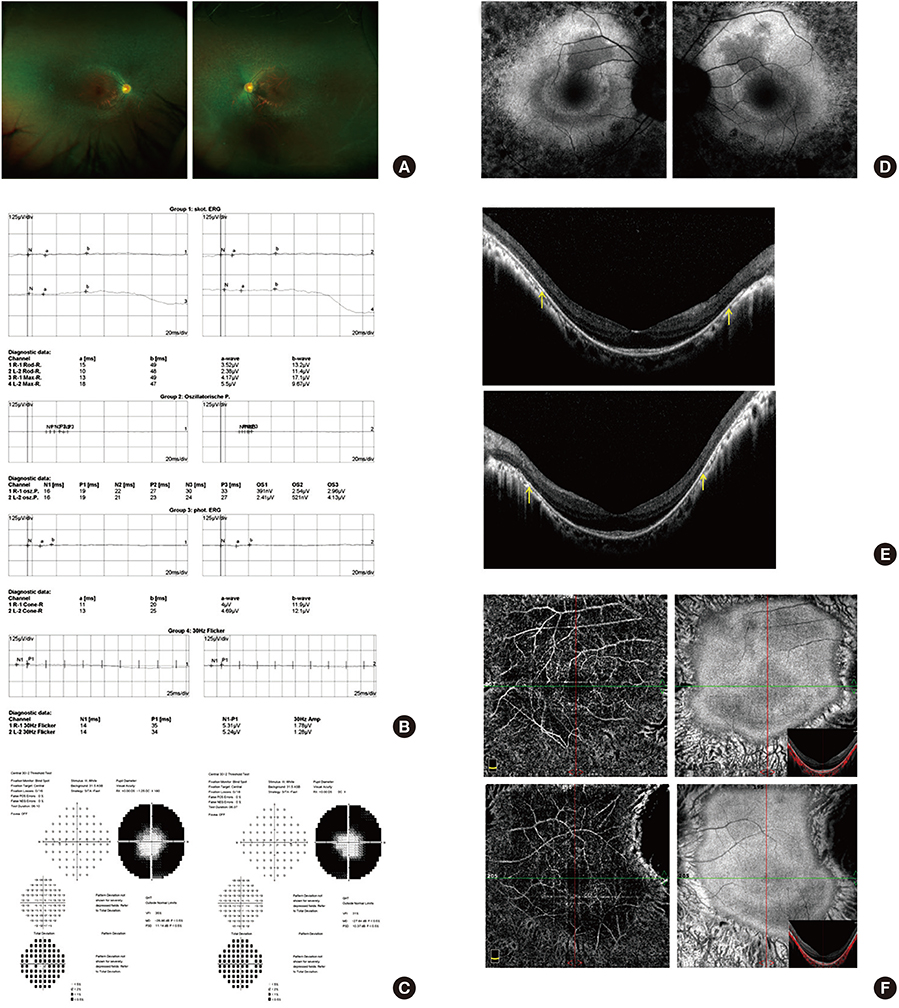
Figure 1 A 35-year-old woman with retinitis pigmentosa. Her best-corrected visual acuity was 20/20 in the right eye and 20/20 in the left eye. (A) Color fundus photographs of both eyes show mottling and granularity at the level of the retinal pigment epithelium (RPE), perivascular bone spicule-shaped pigment deposits in the peripheral retina, and attenuated retinal vessels. The picture on the left is the right eye, and that on the right is the left eye. (B) Full field electroretinography (ERG) reveals near absence of both photopic and scotopic responses of both eyes. The a-wave is derived from the cones and rods of the outer photoreceptor layers. The bwave is derived from the inner retina, predominantly Müller glia and ON-bipolar cells. P2 is the bipolar cell-derived component of the rod-isolated b-wave, and P3 is subtracted from the series of rod responses. The left graph shows the right eye, and the right graph shows the left eye. (C) Visual field testing demonstrates marked peripheral loss with a small residual central visual field in both eyes. The picture on the left is the left eye, and that on the right is the right eye. (D) The lack of signal on fundus autofluorescence imaging (FAF) correlates well with the areas of RPE atrophy. FAF demonstrated a perifoveal ring of increased autofluorescence within the macula with thread-like retinal vessels. The picture on the left is the right eye, and that on the right is the left eye. (E) Spectral domain optical coherence tomography (SD-OCT) shows decreased retinal thickness, particularly in the outer layer: decreased thickness of the outer nuclear layer and loss of the external limiting membrane and the junction between inner segments and outer segments (IS/OS) line (between yellow arrows). The upper picture is the right eye, and the lower is the left eye. (F) En face structural and angiographic imaging with SD-OCT (inset shows a scan plane taken just below the Bruch's membrane) reveals choriocapillaris in the peripheral region in greater detail compared to the central region, with intact outer retina due to increased light penetration in the atrophic peripheral macula. The upper picture is the right eye, and the lower is the left eye.
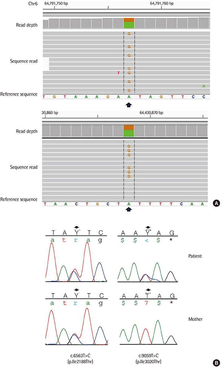
Figure 2 (A) IGV browser visualization of the exome sequencing results of partial genomic DNA sequence of the EYS gene of the patient shows two heterozygous variants. (Upper, c.6563T>C, Lower, c.9059T>C) (B) Validation of EYS variants by Sanger sequencing. The patient had two non-synonymous substitutions—c.[6563T>C];[9059T>C] (arrow)—in EYS. The patient's mother possessed c.6563T>C, but not c.9059T>C.
Cited by 1 articles
-
Genetic Mutation Profiles in Korean Patients with Inherited Retinal Diseases
Min Seok Kim, Kwangsic Joo, Moon-Woo Seong, Man Jin Kim, Kyu Hyung Park, Sung Sup Park, Se Joon Woo
J Korean Med Sci. 2019;34(21):. doi: 10.3346/jkms.2019.34.e161.
Reference
-
1. Hamel C. Retinitis pigmentosa. Orphanet J Rare Dis. 2006; 1:40.
Article2. Abd El-Aziz MM, Barragan I, O'Driscoll CA, Goodstadt L, Prigmore E, Borrego S, et al. EYS, encoding an ortholog of Drosophila spacemaker, is mutated in autosomal recessive retinitis pigmentosa. Nat Genet. 2008; 40:1285–1287.
Article3. Iwanami M, Oshikawa M, Nishida T, Nakadomari S, Kato S. High prevalence of mutations in the EYS gene in Japanese patients with autosomal recessive retinitis pigmentosa. Invest Ophthalmol Vis Sci. 2012; 53:1033–1040.
Article4. Arai Y, Maeda A, Hirami Y, Ishigami C, Kosugi S, Mandai M, et al. Retinitis pigmentosa with EYS mutations is the most prevalent inherited retinal dystrophy in Japanese populations. J Ophthalmol. 2015; 2015:819760.5. Yoon CK. Strategies for mutation discovery in retinitis pigmentosa: transition to the next generation. J Genet Med. 2013; 10:13–19.
Article6. Jang MA, Lee T, Lee J, Cho EH, EH CS. Identification of a novel de novo variant in the PAX3 gene in Waardenburg syndrome by diagnostic exome sequencing: the first molecular diagnosis in Korea. Ann Lab Med. 2015; 35:362–365.
Article7. Kumar P, Henikoff S, Ng PC. Predicting the effects of coding non-synonymous variants on protein function using the SIFT algorithm. Nat Protoc. 2009; 4:1073–1081.
Article8. Adzhubei IA, Schmidt S, Peshkin L, Ramensky VE, Gerasimova A, Bork P, et al. A method and server for predicting damaging missense mutations. Nat Methods. 2010; 7:248–249.
Article9. Matsuya A, Sakate R, Kawahara Y, Koyanagi KO, Sato Y, Fujii Y, et al. Evola: Ortholog database of all human genes in H-InvDB with manual curation of phylogenetic trees. Nucleic Acids Res. 2008; 36:D787–D792.
Article10. Di Y, Huang L, Sundaresan P, Li S, Kim R, Ballav Saikia B, et al. Whole-exome sequencing analysis identifies mutations in the EYS gene in retinitis pigmentosa in the Indian population. Sci Rep. 2016; 6:19432.
Article11. Gurudev N, Yuan M, Knust E. chaoptin, prominin, eyes shut and crumbs form a genetic network controlling the apical compartment of Drosophila photoreceptor cells. Biol Open. 2014; 3:332–341.
Article12. Khan MI, Collin RW, Arimadyo K, Micheal S, Azam M, Qureshi N, et al. Missense mutations at homologous positions in the fourth and fifth laminin A G-like domains of eyes shut homolog cause autosomal recessive retinitis pigmentosa. Mol Vis. 2010; 16:2753–2759.13. Lee SH, Yu HG, Seo JM, Moon SW, Moon JW, Kim SJ, et al. Hereditary and clinical features of retinitis pigmentosa in Koreans. J Korean Med Sci. 2010; 25:918–923.
Article
- Full Text Links
-
- Actions
-
Cited
- CITED
-
- Close
- Share
-
- Similar articles
-
- Genetic Characteristics of 63 Patients with Non-syndromic Retinitis Pigmentosa at a Single Korean Institution
- A Case of Retinitis Pigmentosa without Pigment
- A Case of Unilateral Retinitis Pigmentosa
- Strategies for Mutation Discovery in Retinitis Pigmentosa: Transition to the Next Generation
- Visual Function and Functional Vision of Retinitis Pigmentosa
