Imaging Sci Dent.
2018 Mar;48(1):59-65. 10.5624/isd.2018.48.1.59.
Unusual malignant neoplasms occurring around dental implants: A report of 2 cases
- Affiliations
-
- 1Department of Oral and Maxillofacial Radiology, Graduate School, Kyung Hee University, Seoul, Korea. omrcys@khu.ac.kr
- KMID: 2406984
- DOI: http://doi.org/10.5624/isd.2018.48.1.59
Abstract
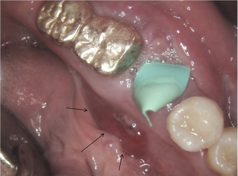
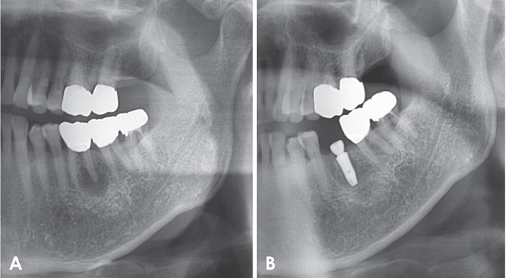
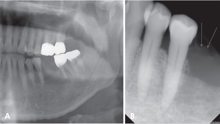
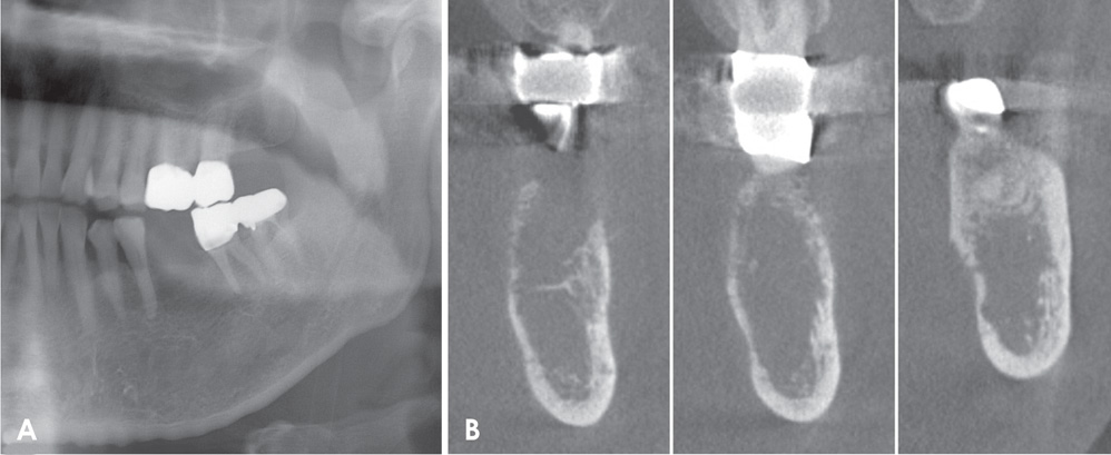
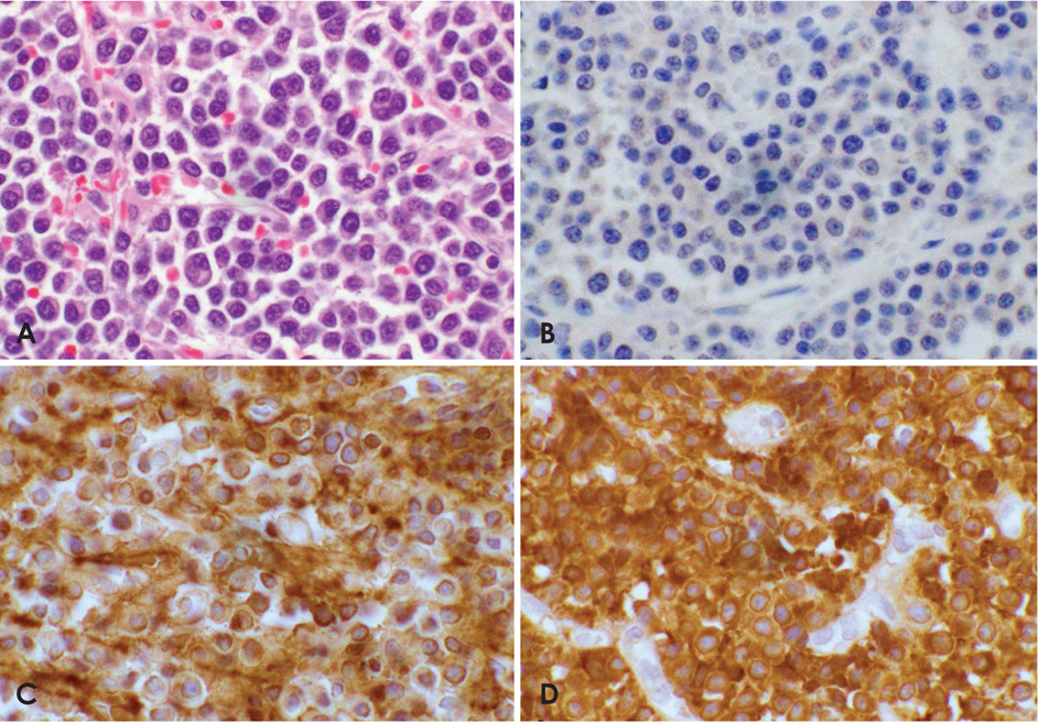
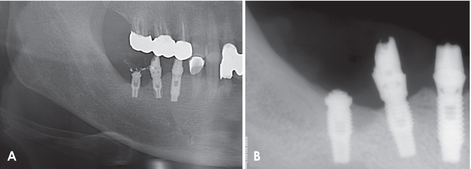
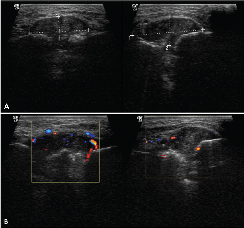
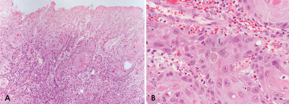
- Osseointegrated implants are now commonplace in contemporary dentistry. However, a number of complications can occur around dental implants, including peri-implantitis, maxillary sinusitis, osteomyelitis, and neoplasms. There have been several reports of a malignant neoplasm occurring adjacent to a dental implant. In this report, we describe 2 such cases. One case was that of a 75-year-old man with no previous history of malignant disease who developed a solitary plasmacytoma around a dental implant in the left posterior mandible, and the other was that of a 43-year-old man who was diagnosed with squamous cell carcinoma adjacent to a dental implant in the right posterior mandible. Our experiences with these 2 cases suggest the possibility of a relationship between implant treatment and an inflammatory cofactor that might increase the risk of development of a malignant neoplasm.
MeSH Terms
Figure
Reference
-
1. Henry PJ. Oral implant restoration for enhanced oral function. Clin Exp Pharmacol Physiol. 2005; 32:123–127.
Article2. Atieh MA, Alsabeeha NH, Faggion CM Jr, Duncan WJ. The frequency of peri-implant diseases: a systematic review and meta-analysis. J Periodont. 2013; 84:1586–1598.
Article3. McDermott NE, Chuang SK, Woo VV, Dodson TB. Complications of dental implants: identification, frequency, and associated risk factors. Int J Oral Maxillofac Implants. 2003; 18:848–855.
Article4. Poggio CE. Plasmacytoma of the mandible associated with a dental implant failure: a clinical report. Clin Oral Implants Res. 2007; 18:540–543.
Article5. Junquera L, Gallego L, Pelaz A. Multiple myeloma and bisphosphonate-related osteonecrosis of the mandible associated with dental implants. Case Rep Dent. 2011; 2011:568246.
Article6. Rahal MD, Brånemark PI, Osmond DG. Response of bone marrow to titanium implants: osseointegration and the establishment of a bone marrow-titanium interface in mice. Int J Oral Maxillofac Implants. 1993; 8:573–579.7. Fulop GM, Osmond DG. Regulation of bone marrow lymphocyte production. III. Increased production of B and non-B lymphocytes after administering systemic antigens. Cell Immunol. 1983; 75:80–90.8. Pietrangeli CE, Osmond DG. Regulation of B-lymphocyte production in the bone marrow: mediation of the effects of exogenous stimulants by adoptively transferred spleen cells. Cell Immunol. 1987; 107:348–357.
Article9. Osmond DG, Priddle S, Rico-Vargas S. Proliferation of B cell precursors in bone marrow of pristane-conditioned and malaria-infected mice: implications for B cell oncogenesis. Curr Top Microbiol Immunol. 1990; 166:149–157.
Article10. Hussein MA, George R, Rybicki L, Karam MA. Skeletal trauma preceding the development of plasma cell dyscrasia: eight case reports and review of the literature. Med Oncol. 2003; 20:349–354.
Article11. Caligaris-Cappio F, Bergui L, Gregoretti MG, Gaidano G, Gaboli M, Schena M, et al. Role of bone marrow stromal cells in the growth of human multiple myeloma. Blood. 1991; 77:2688–2693.
Article12. McGrory JE, Pritchard DJ, Unni KK, Ilstrup D, Rowland CM. Malignant lesions arising in chronic osteomyelitis. Clin Orthop Relat Res. 1999; 362:181–189.
Article13. Oehler R, Weingartmann G, Manhart N, Salzer U, Meissner M, Schlegel W, et al. Polytrauma induces increased expression of pyruvate kinase in neutrophils. Blood. 2000; 95:1086–1092.
Article14. Block MS, Scheufler E. Squamous cell carcinoma appearing as peri-implant bone loss: a case report. J Oral Maxillofac Surg. 2001; 59:1349–1352.
Article15. Czerninski R, Kaplan I, Almoznino G, Maly A, Regev E. Oral squamous cell carcinoma around dental implants. Quintessence Int. 2006; 37:707–711.16. Eguia del Valle A, Martinez-Conde Llamosas R, López Vicente J, Uribarri Etxebarria A, Aguirre Urizar JM. Primary oral squamous cell carcinoma arising around dental osseointegrated implants mimicking peri-implantitis. Med Oral Patol Oral Cir Bucal. 2008; 13:E489–E491.17. Schache A, Thavaraj S, Kalavrezos N. Osseointegrated implants: a potential route of entry for squamous cell carcinoma of the mandible. Br J Oral Maxillofac Surg. 2008; 46:397–399.
Article
- Full Text Links
- Actions
-
Cited
- CITED
-
- Close
- Share
- Similar articles
-
- Disappearance of a dental implant after migration into the maxillary sinus: an unusual case
- Tissue Engineering for Dental Implants
- A stress analysis of fixed prostheses with dental implant and natural tooth
- A study on the effects of early loading on the surrounding bone tissue of the dental implants
- A study on the effect of rotational speeds of the trephine mill on the temperature of surrounding bone during dental implantation procedure and osseointegration of implants

