Lesions that Mimic Musculoskeletal Infection: A Pictorial Essay
- Affiliations
-
- 1Department of Radiology, Kyung Hee University Hospital, College of Medicine, Kyung Hee University, Seoul, Korea. t2star@naver.com
- 2Department of Radiology, Kyung Hee University Hospital at Gangdong, College of Medicine, Kyung Hee University, Seoul, Korea.
- KMID: 2405723
- DOI: http://doi.org/10.3348/jksr.2018.78.3.200
Abstract
- Musculoskeletal (MSK) infections, such as osteomyelitis, infectious arthritis and spondylitis have variable radiographic findings depending on their underlying cause and clinical infection stage. Other disease entities, ranging from simple degenerative lesions to tumorous bone conditions, in which there is no evidence of infectious origin, can share similar radiographic findings. It is important to be aware, when interpreting radiographic features that are typically associated with MSK infections, that a non-infectious MSK disease may be mimicking the radiographic findings of infectious diseases.
MeSH Terms
Figure
-
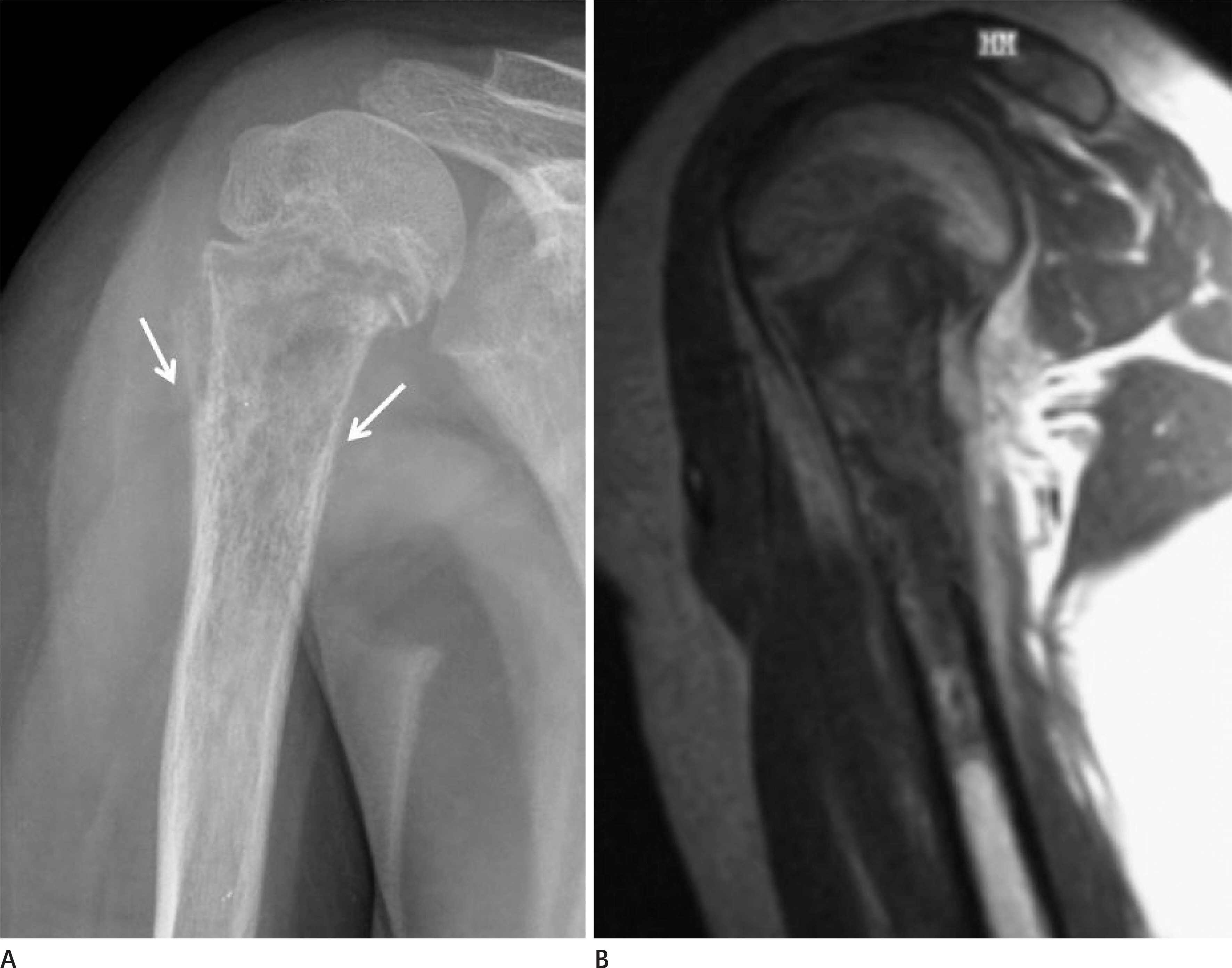
Fig. 1. A 10-year-old female with osteosarcoma. A. Proximal metaphysis of the right humerus shows “moth-eaten” and permeative bone destruction (arrows). B. On contrast-enhanced T1-weighted MRI, strong enhancement of the soft tissue adjacent to the destructive bone lesion is evident, suggesting inflammation.
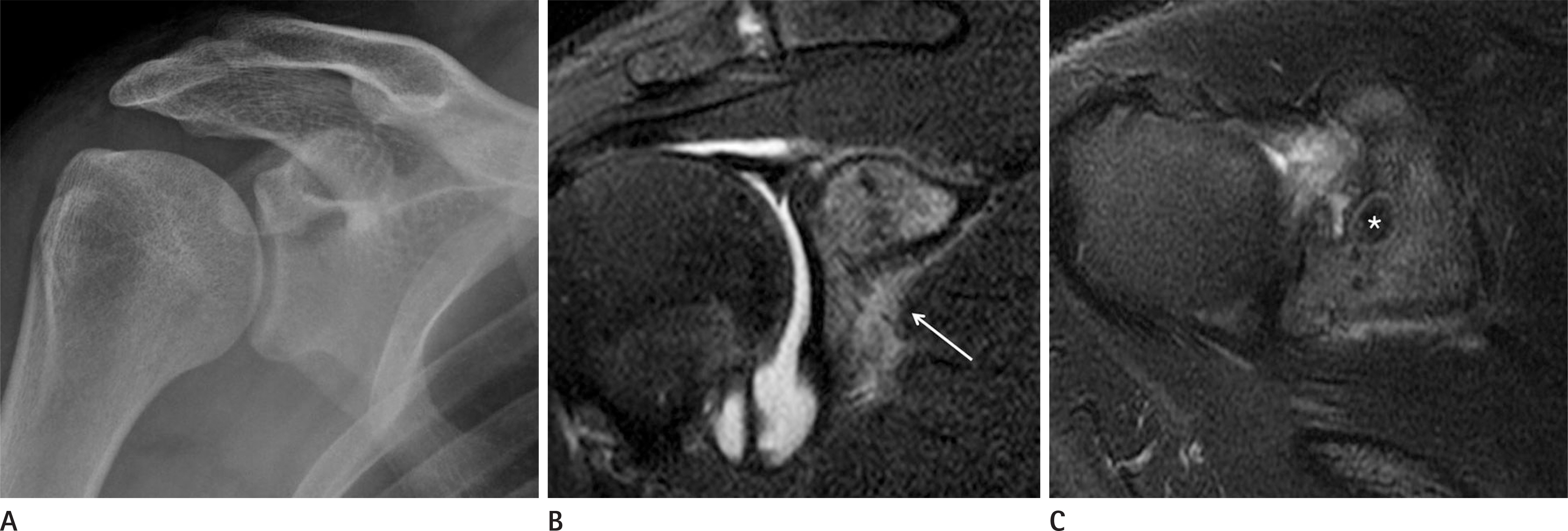
Fig. 2. A 21-year-old male with osteoid osteoma. A. Plain radiograph of the right shoulder shows normal findings. B. On coronal T2 fat suppression MRI, the high signal intensity of the coracoid process is indicative of bone marrow edema. Inflammatory changes of the adjacent soft tissues are also present (arrow). C. Axial T2 fat suppression MRI taken one year later shows a well-circumscribed round mass-like lesion (asterisk) at the base of the coracoid process. It appears as a low signal intensity lesion with a high-signal-intensity peripheral rim. Reactive sclerosis and bone marrow edema have developed around the lesion.
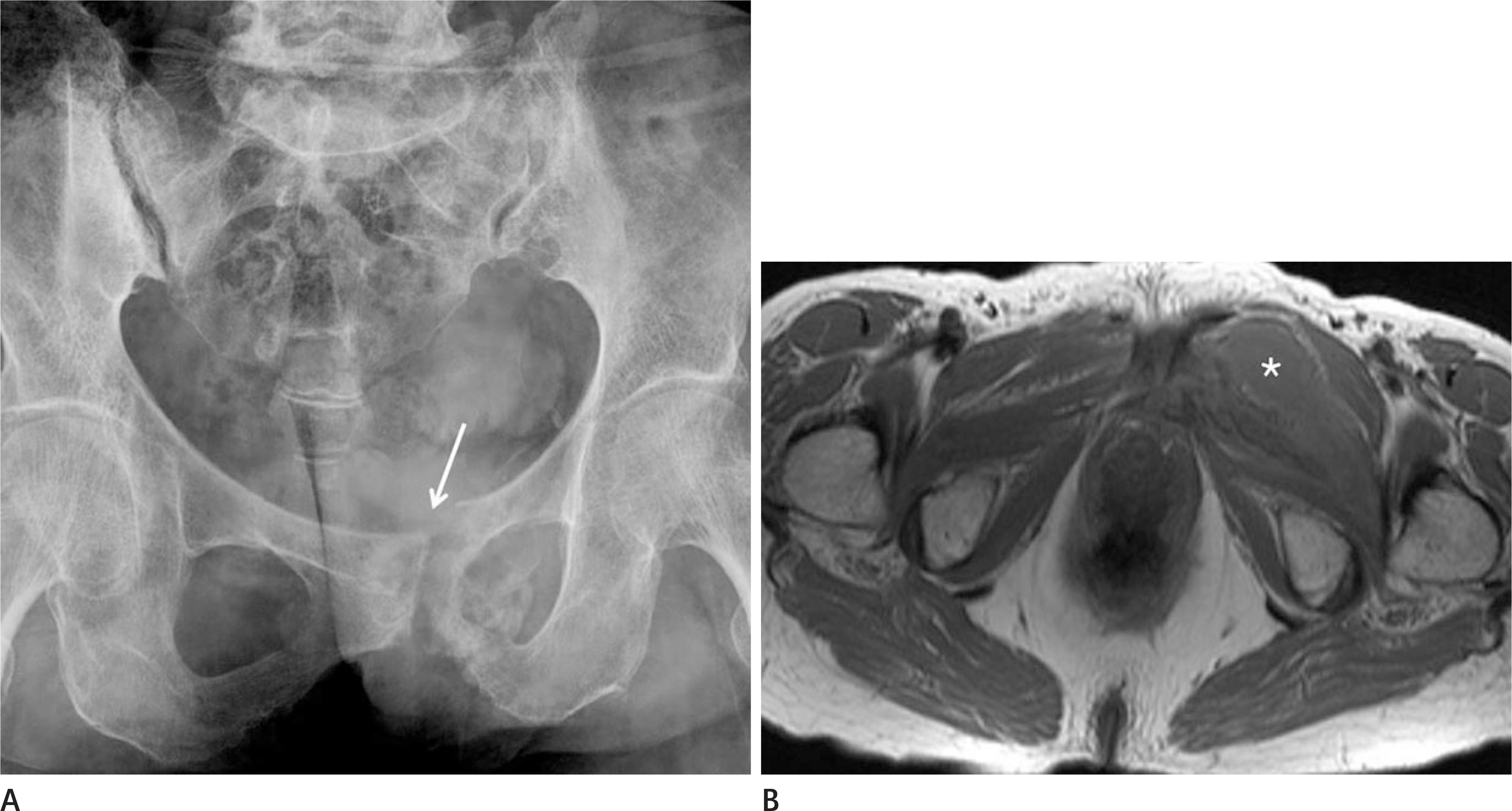
Fig. 3. A 65-year-old female with insufficiency fracture of the pelvis. A. Plain radiograph of the pelvis shows a left pubic bone fracture (arrow) with reactive sclerosis and bone formation. B. Axial contrast-enhanced MRI shows extensive perilesional bone marrow signal changes. Also, there is localized fluid collection that seems to be due to a hematoma (asterisk) near the fracture site. Such findings can easily be mistaken for an abscess pocket.
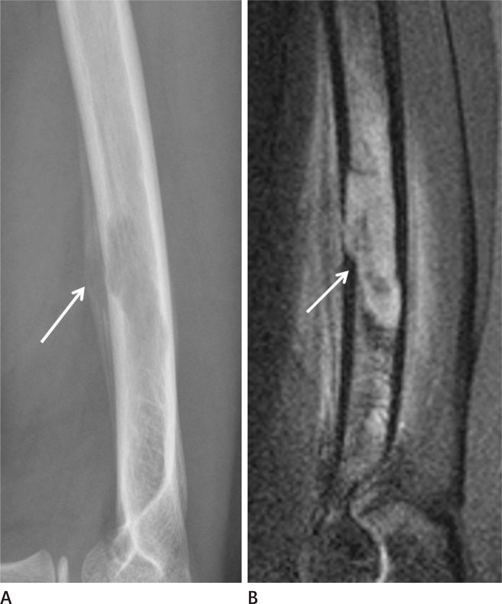
Fig. 4. A 10-year-old male with Langerhan's cell histiocytosis. A. Plain radiograph of the humerus shows a lobulated osteolytic lesion with periosteal reaction (arrow) at the mid shaft. There is no evidence of perilesional reactive sclerosis. B. Contrast-enhanced MRI in the sagittal plane shows osteolytic lesions with adjacent cortical destruction and periosteal reaction (arrow). Swelling and enhancement of the surrounding soft tissues are also visible.
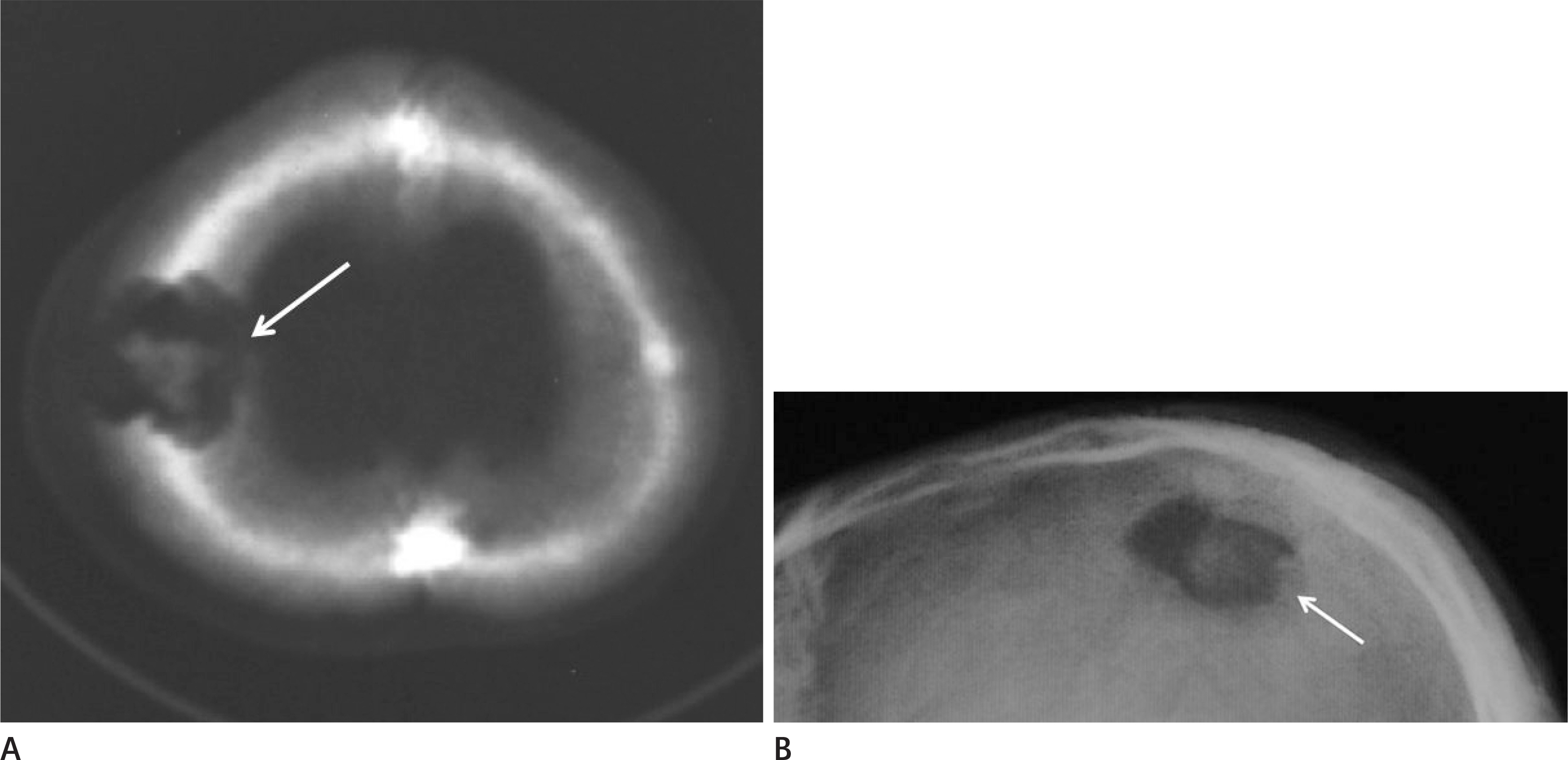
Fig. 5. A 19-year-old male with Langerhan's cell histiocytosis. A. An axial CT image of the skull shows an osteolytic lesion with a lobulated contour (arrow). At the center of the osteolytic lesion, there is a bony lesion with an irregular margin that is indicative of a sequestrum. B. Similarly, the plain radiograph of the same patient's skull shows an osteolytic lesion (arrow) with an inner high-density portion. However, neither reactive sclerosis nor significant periosteal reactions are apparent near the osteolytic lesion.
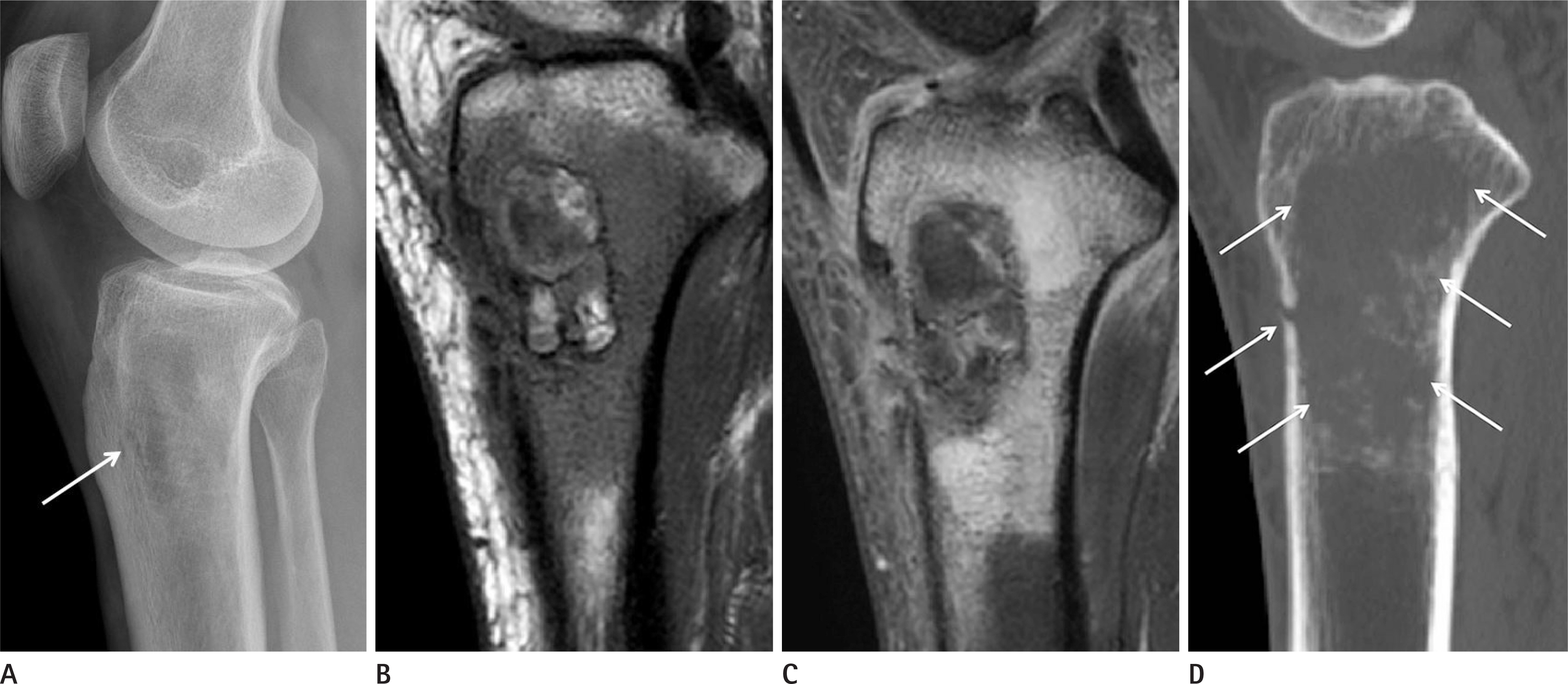
Fig. 6. A 58-years-old female with fibroblastic osteosarcoma, grade IV. A. Plain radiograph shows an ill-defined osteolytic bone lesion (arrow) accompanied by minimal sclerosis at the metaphysis of the left proximal tibia. There is no periosteal reaction. B, C. On sagittal T1-weighted (B) and contrast-enhanced T1-spectral presaturation with inversion recovery MRI (C), the bony destructive lesion has a lobulated contour with inner septa-like structures. There is an inner rim of intermediate to high signal intensity, and an outer rim of dark signal intensity. D. CT image of the tibia shows more extensive bone destruction (arrows) than what was observed from MR images. Also, the low signal rim seen in MRI was not a bony sclerosis.
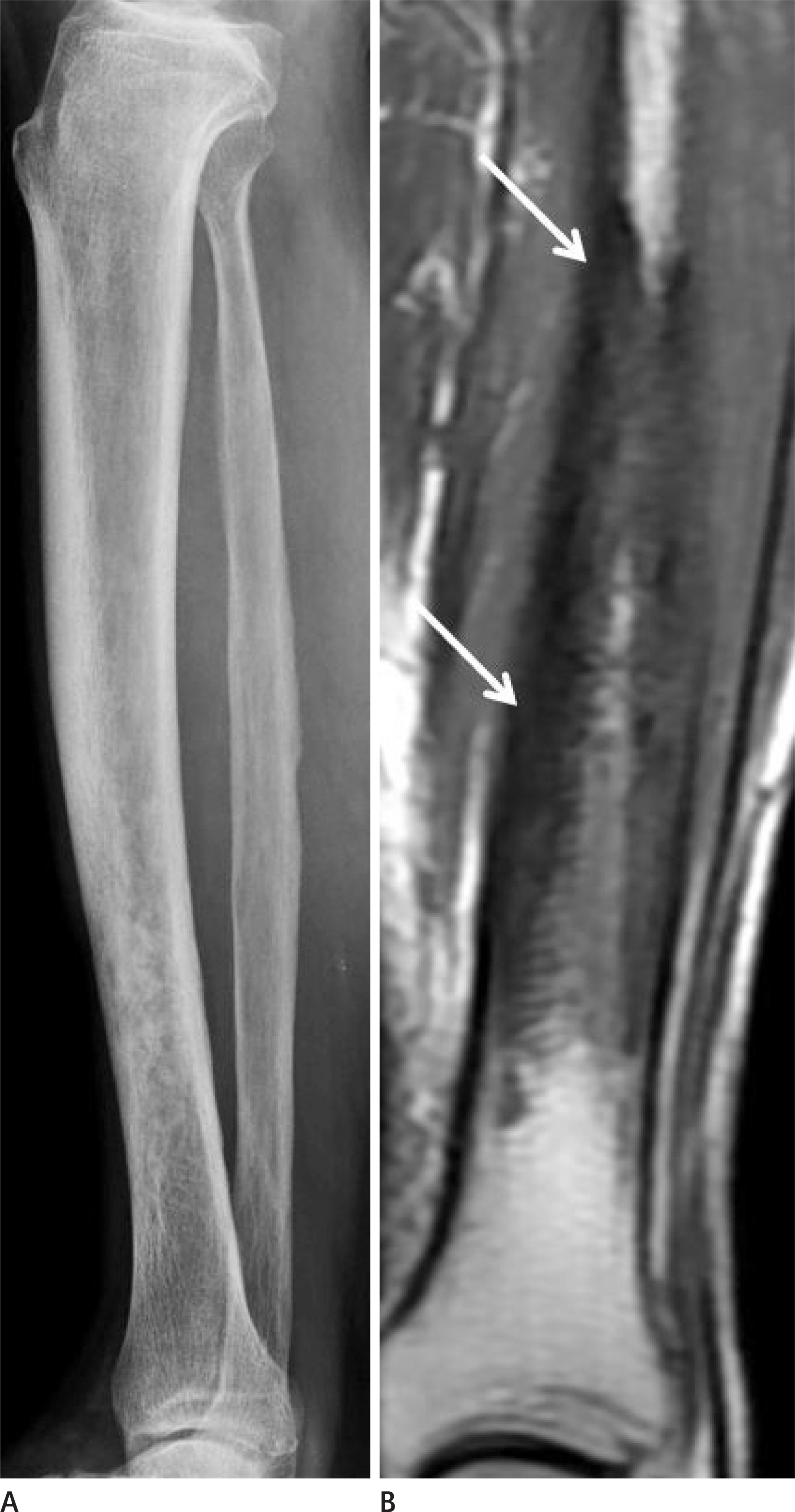
Fig. 7. A 73-year-old male with bone metastasis. A. Plain radiograph of the left lower extremity shows irregular cortical thickening with extensive sclerosis along the distal shaft of the tibia. B. T1-weighted MRI in the coronal plane shows diffuse thickening of the cortex with increased signal intensity. Also, the bone marrow signal intensity is darker than normal (arrows).
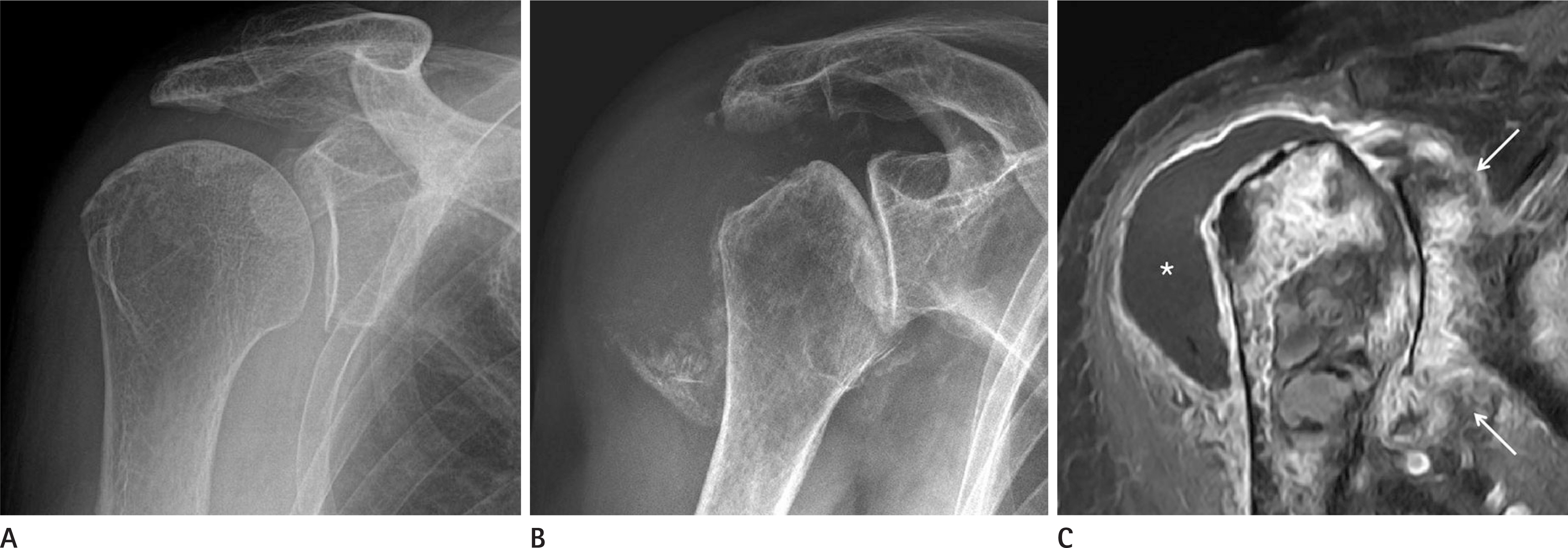
Fig. 8. An 80-year-old female with neurotrophic arthropathy. A. Plain radiograph of the patient's right shoulder shows only mild degenerative changes without joint space narrowing. B. After 8 months, her right shoulder joint shows rapid destruction of its articular margins. Multifocal detached osseous fragments are visible. C. Contrast-enhanced T1-spectral presaturation with inversion recovery MRI in the coronal plane shows diffuse enhancement along the subchondral portion and the joint capsule (arrows). Also, capsular distension caused by a large amount of effusion (asterisk) can be seen. However, periarticular soft tissue changes are not prominent.
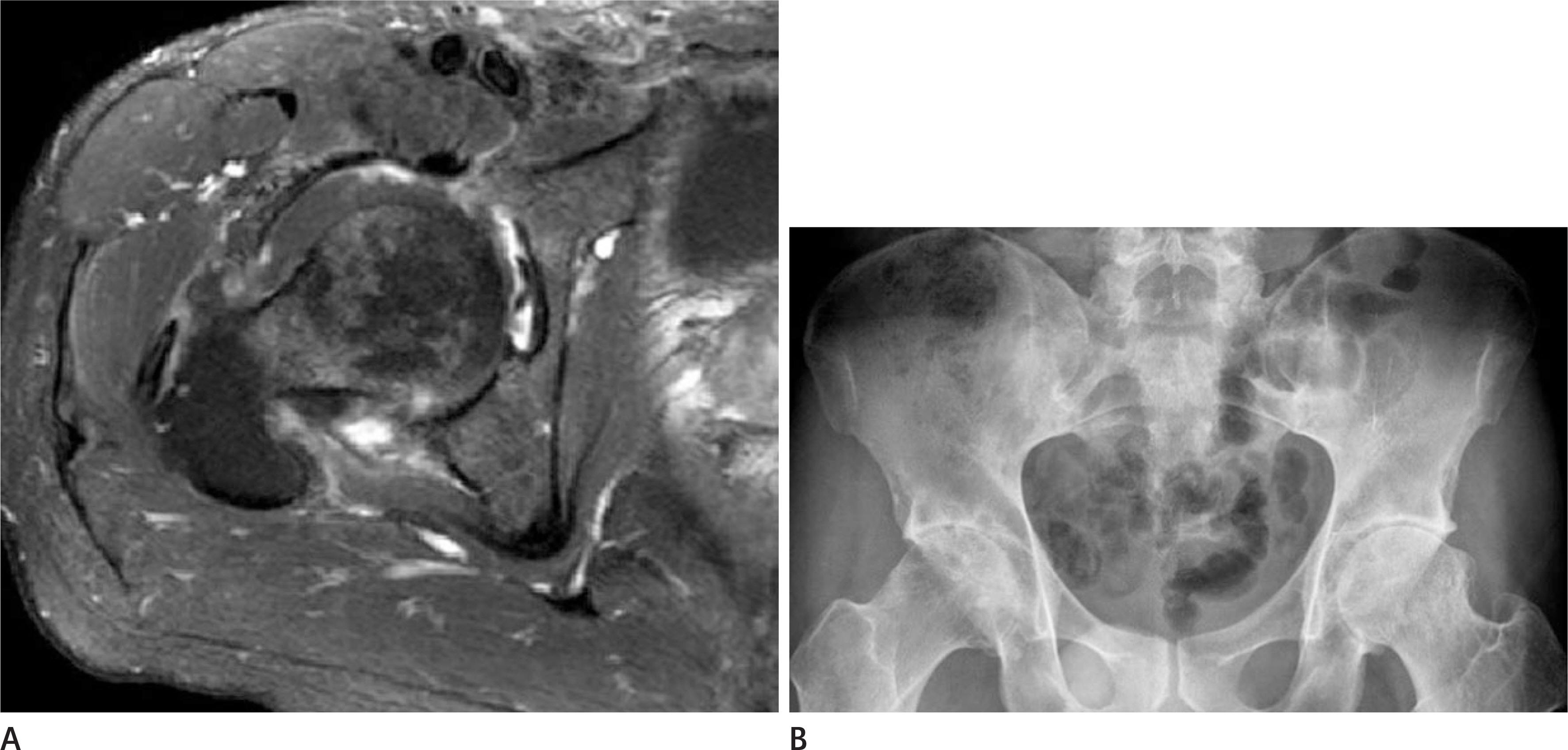
Fig. 9. A 35-year-old male with ankylosing spondylitis involvement of hip joints. A. Axial contrast-enhanced T1-spectral presaturation with inversion recovery MRI of the right hip joint shows periarticular soft tissue edema. Joint effusion with capsular distension and the narrowing of the joint space are also evident. However, there are no significant signal changes in the bony structures. B. Plain radiograph of the pelvis in the same patient shows complete ankylosis of both sacroiliac joints. This patient has an underlying ankylosing spondylitis with involvement of both hip joints.
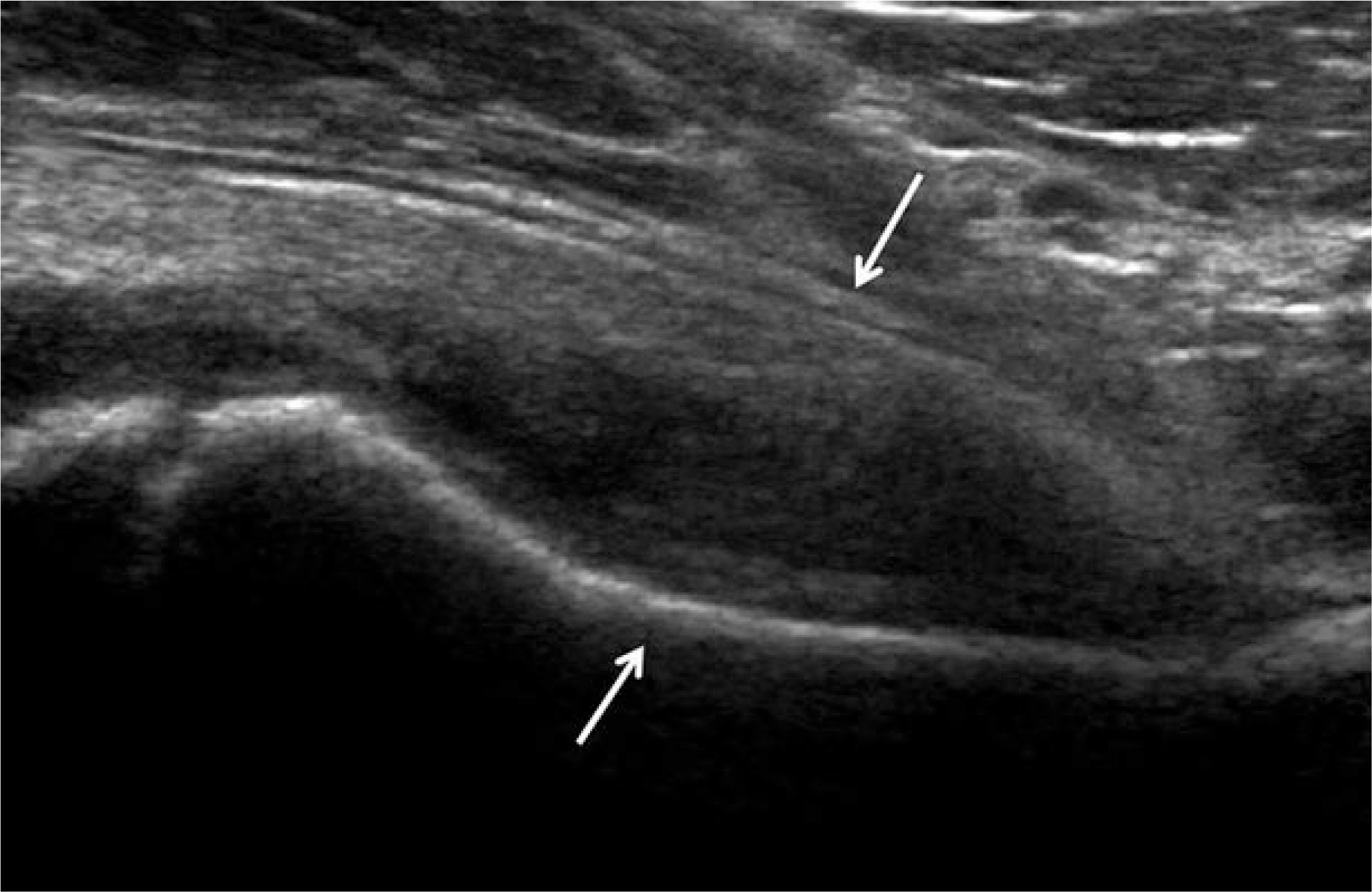
Fig. 10. An 8-year-old female with transient synovitis of the hip. Ultrasound findings of the right hip joint include joint effusion with synovial hypertrophy and capsular distention (arrows). However, the capsular margin is well preserved and there are few pericapsular changes.
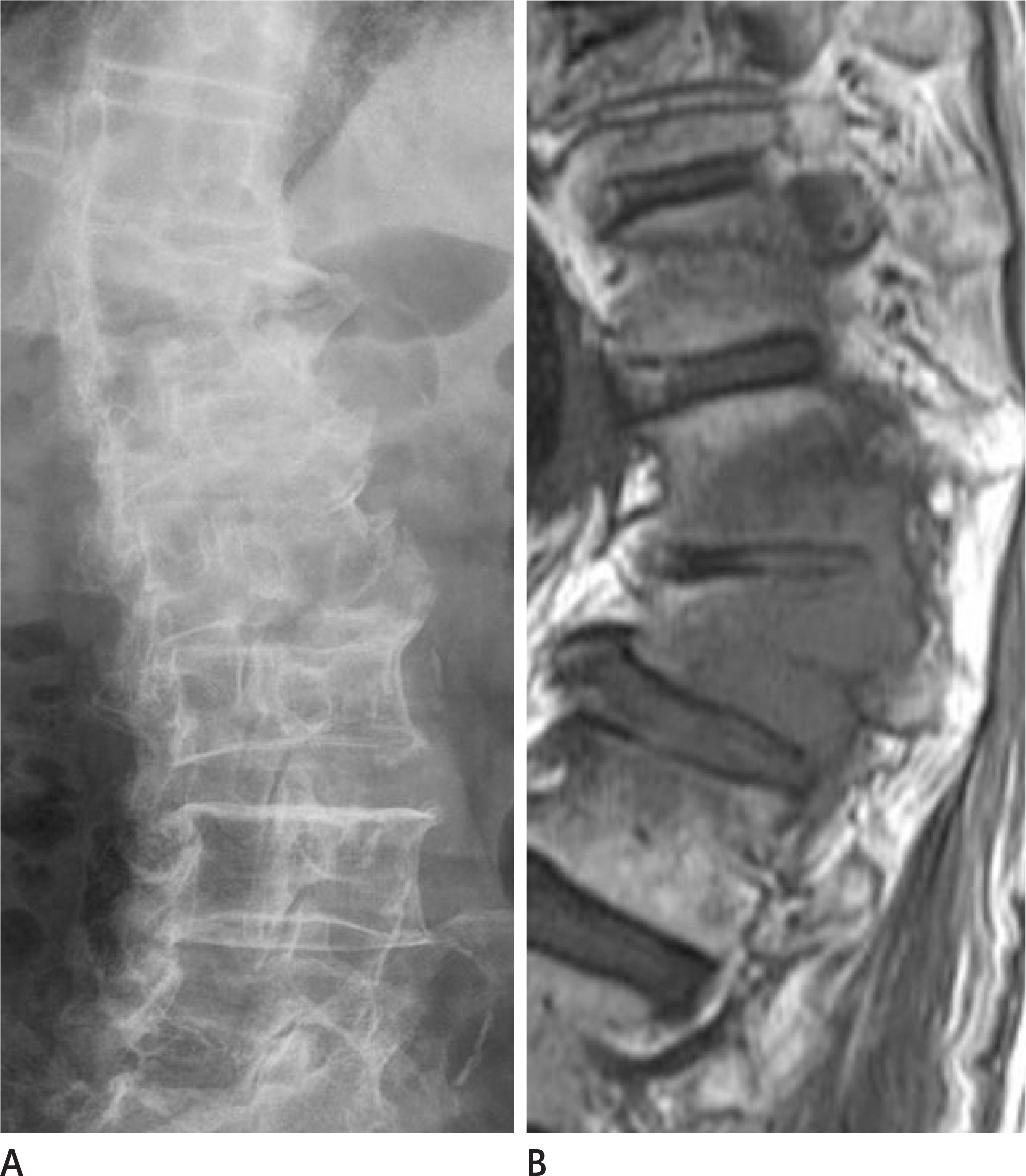
Fig. 11. A 68-year-old male with vertebral metastasis. A. Plain radiograph of T-L spine in the lateral view shows bony ankylosis at the T10-T12 levels. This finding is suggestive of sequelae of infectious spondylitis. B. Sagittal T1-weighted MRI shows decreased body heights of T11 and T12 as well as partial ankylosis. The right lateral portions and the pedi-cles are seen as low signal intensity and there is also a paravertebral mass formation on the right side.
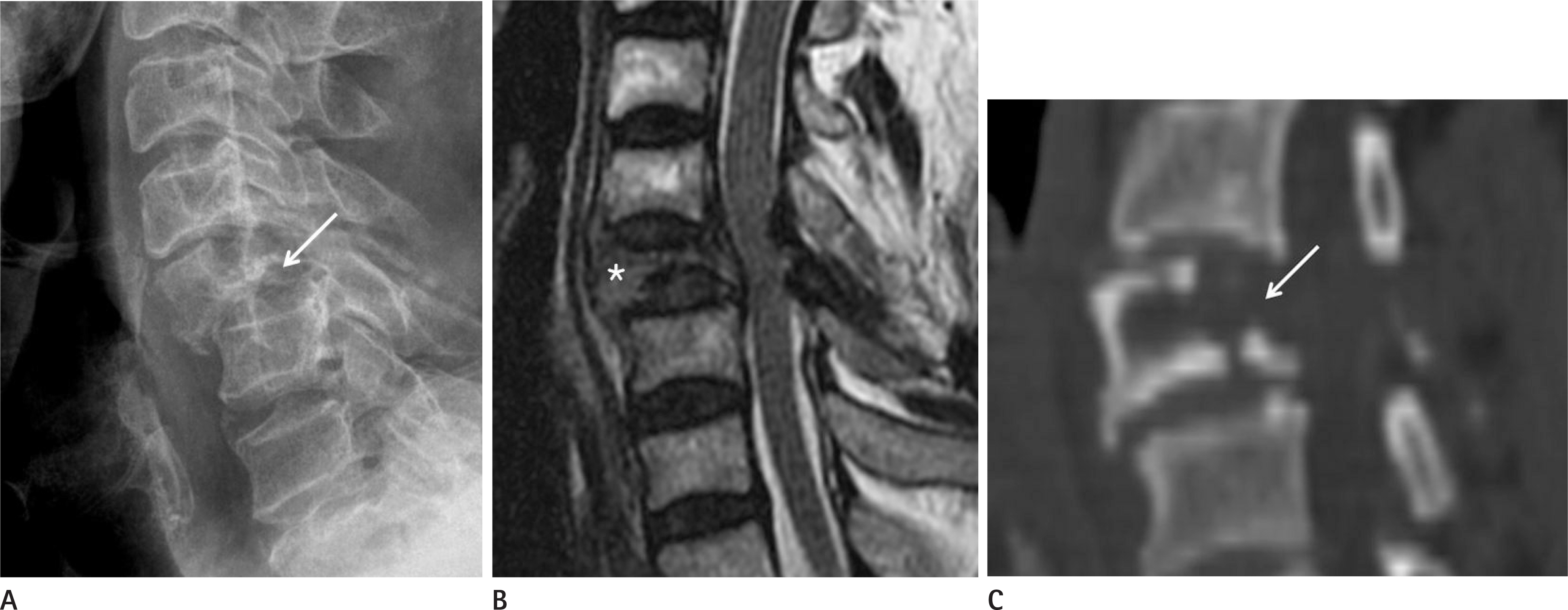
Fig. 12. A 73-year-old male with vertebral metastasis. A. Plain radiograph of the C-spine in the lateral view shows a collapsed C5 body with severe anterolisthesis upon C6 (arrow). B. Sagittal T2-weighted MRI of the C-spine shows a pathologic compression fracture at the C5 body (asterisk) as well as an epidural mass formation. C. Sagittal CT image of the same patient shows osteolytic bone destruction with reactive sclerosis on the remaining bones (arrow). Such findings may be seen in infectious lesions like tuberculosis.
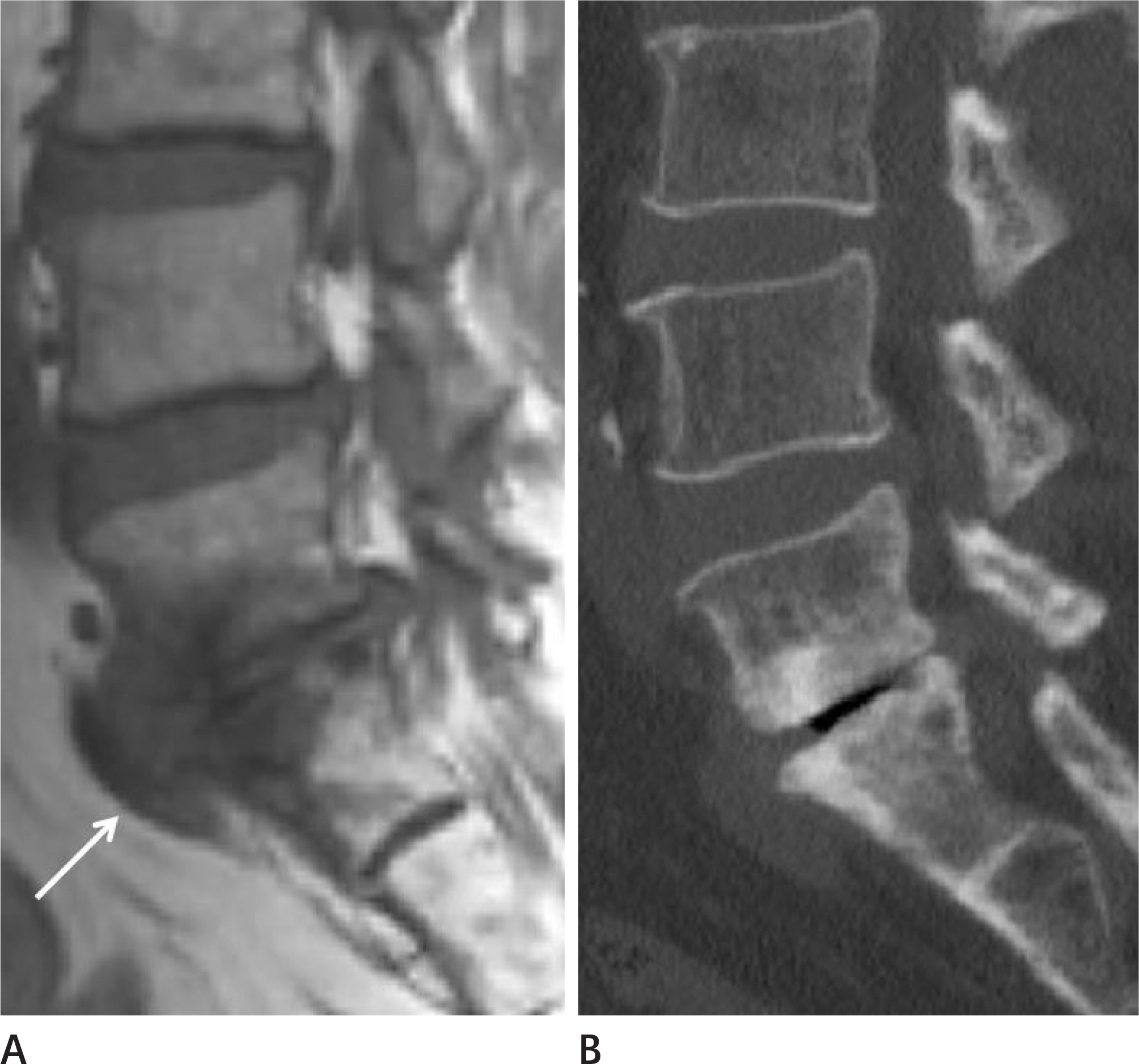
Fig. 13. A 60-year-old male with degenerative changes of L-spine. A. Sagittal T2-weighted MRI of the L-spine shows severe disc degeneration with bone erosion (arrow) caused by mechanical forces at L5-S1. B. Sagittal CT image of the same patient shows disc herniation at L5-S1, with secondary sclerotic changes in the adjacent vertebral bodies.
Reference
-
References
1. Poultsides LA, Liaropoulos LL, Malizos KN. The socioeconomic impact of musculoskeletal infections. J Bone Joint Surg Am. 2010; 92:e13.
Article2. Manaster BJ. Diagnostic imaging: musculoskeletal non-traumatic disease. Philadelphia, PA: Elsevier Health Sciences;2016.3. Resnick D. Osteomyelitis, septic arthritis, and soft tissue infection: organisms. In Resnick D, ed. Bone and Joint Imaging. Philadelphia, PA: Saunders;2006. p. 753–788.4. Pineda C, Espinosa R, Pena A. Radiographic imaging in osteomyelitis: the role of plain radiography, computed tomography, ultrasonography, magnetic resonance imaging, and scintigraphy. Semin Plast Surg. 2009; 23:80–89.
Article5. Peh WC, Khong PL, Yin Y, Ho WY, Evans NS, Gilula LA, et al. Imaging of pelvic insufficiency fractures. Radiographics. 1996; 16:335–348.
Article6. Hayes CS, Heinrich SD, Craver R, MacEwen GD. Subacute osteomyelitis. Orthopedics. 1990; 13:363–366.
Article7. Mhuircheartaigh JN, Lin YC, Wu JS. Bone tumor mimickers: a pictorial essay. Indian J Radiol Imaging. 2014; 24:225–236.
Article8. Khung S, Budzik JF, Amzallag-Bellenger E, Lambilliote A, Soto Ares G, Cotten A, et al. Skeletal involvement in Langerhans cell histiocytosis. Insights Imaging. 2013; 4:569–579.
Article9. Kaul R, Gupta N, Gupta S, Gupta M. Eosinophilic granuloma of skull bone. J Cytol. 2009; 26:156–157.
Article10. Joo I, Choi JA, Chung JH, Oh JH, Hong SH, Kang HS. Fibroblastic type osteosarcoma of the ulna: a case report of a tumor in a rare location with atypical imaging findings. Korean J Radiol. 2009; 10:85–88.
Article11. Sundaram M, Totty WG, Kyriakos M, McDonald DJ, Merkel K. Imaging findings in pseudocystic osteosarcoma. AJR Am J Roentgenol. 2001; 176:783–788.
Article12. Perron AD, Brady WJ, Miller MD. Orthopedic pitfalls in the ED: osteomyelitis. Am J Emerg Med. 2003; 21:61–67.
Article13. Khanna G, Sato TSP, Ferguson P. Imaging of chronic recurrent multifocal osteomyelitis. Radiographics. 2009; 29:1159–1177.
Article14. Giurato L, Uccioli L. The diabetic foot: Charcot joint and osteomyelitis. Nucl Med Commun. 2006; 27:745–749.
Article15. Vander Cruyssen B, Muñoz-Gomariz E, Font P, Mulero J, de Vlam K, Boonen A, et al. ASPECT-REGISPONSER-RESPON-DIA working group. Hip involvement in ankylosing spondylitis: epidemiology and risk factors associated with hip replacement surgery. Rheumatology (Oxford). 2010; 49:73–81.
Article16. Jeong H, Eun YH, Kim IY, Kim H, Lee J, Koh EM, et al. Characteristics of hip involvement in patients with ankylosing spondylitis in Korea. Korean J Intern Med. 2017; 32:158–164.
Article17. Hong SH, Choi JY, Lee JW, Kim NR, Choi JA, Kang HS. MR imaging assessment of the spine: infection or an imitation? Radiographics. 2009; 29:599–612.
Article18. Shah LM, Salzman KL. Imaging of spinal metastatic disease. Int J Surg Oncol. 2011; 2011:769753.
Article19. Khattry N, Thulkar S, Das A, Khan SA, Bakhshi S. Spinal tuberculosis mimicking malignancy: atypical imaging features. Indian J Pediatr. 2007; 74:297–298.
Article
- Full Text Links
-
- Actions
-
Cited
- CITED
-
- Close
- Share
-
- Similar articles
-
- Musculoskeletal Applications of Elastography: a Pictorial Essay of Our Initial Experience
- RE: Musculoskeletal Applications of Elastography: a Pictorial Essay of Our Initial Experience
- Incidental Musculoskeletal Lesions Detected on Abdominopelvic CT Scans: A Pictorial Essay
- Imaging Features of the Mesenchymal Tumors of the Breast according to WHO Classification: A Pictorial Essay
- Breast lesions during pregnancy and lactation: a pictorial essay
