Extrinsic Compression of the Left Main Coronary Artery by a Huge Aortic Arch Aneurysm Mimicking Acute Coronary Syndrome
- Affiliations
-
- 1Department of Cardiology, Ajou University School of Medicine, Suwon, Korea. sychoimd@outlook.com
- 2Department of Cardiac Surgery, Ajou University School of Medicine, Suwon, Korea.
- 3Department of Radiology, Ajou University School of Medicine, Suwon, Korea.
- KMID: 2405053
- DOI: http://doi.org/10.4070/kcj.2017.0313
Abstract
- No abstract available.
Figure
-
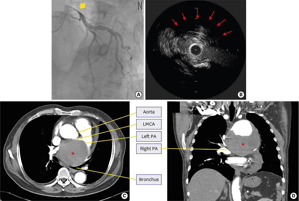
Figure 1 Findings of coronary angiography, IVUS and chest CTA. (A) Coronary angiography showing a bird beak-like significant narrowing of the ostium of the LMCA (yellow bold arrow). (B) IVUS demonstrated that the lumen of the LMCA ostium was deformed as a fusiform narrowing with mild atherosclerotic plaque surrounded by decreased echo-signals behind the vessel suspected of extrinsic compression (red arrows). (C, D) Immediate chest CTA revealed a huge 10×11 cm sized aortic arch aneurysm containing a mural thrombus (red asterisk) severely compressed right PA, left PA, left main-stem bronchus, and LMCA. CTA = computed tomographic angiogram; IVUS = intravascular ultrasound; LMCA = left main coronary artery; PA = pulmonary artery.
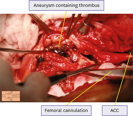
Figure 2 Surgical finding. Image taken during surgery revealing the giant aortic aneurysm containing a red thrombus. ACC = aortic cross clamp.
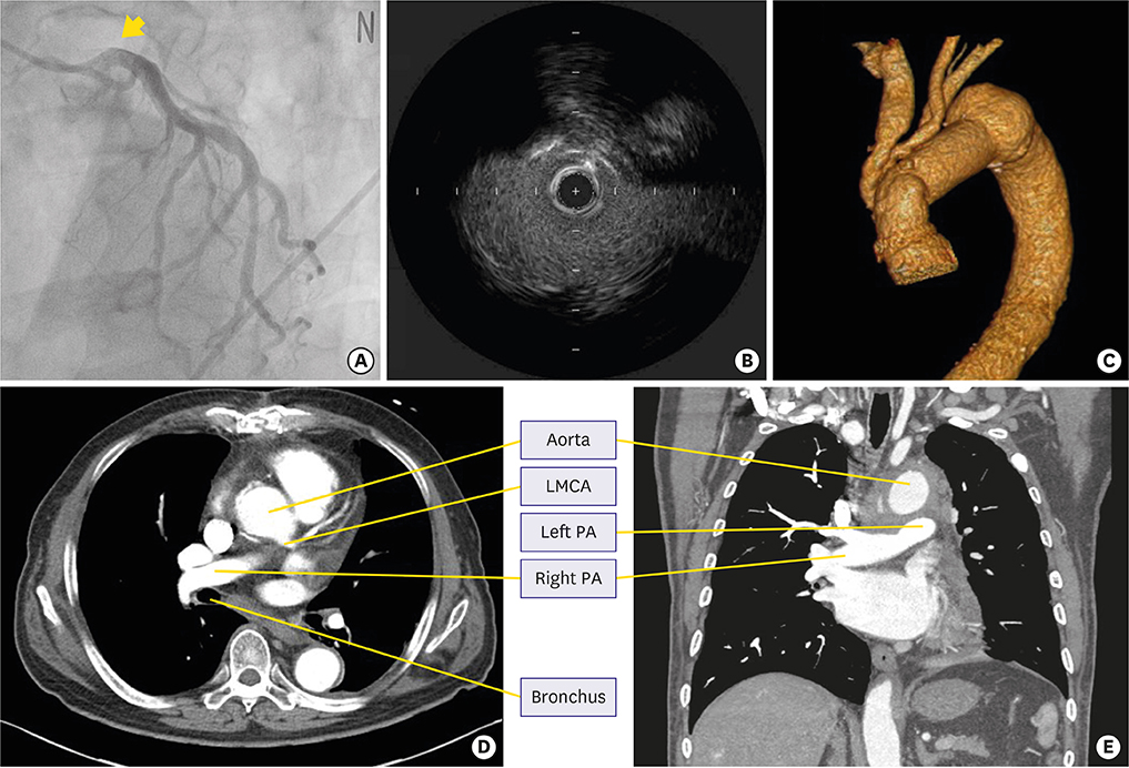
Figure 3 Findings of coronary angiography, IVUS and chest CTA after 4-vessel graft interposition. (A) After ascending aortic arch excision and graft interposition, coronary angiogram shows normal-looking appearance of the left coronary artery ostium. (B) IVUS shows the LMCA lumen was patent with minimal atherosclerotic plaque. (C-E) CTA shows significantly decreased size of aorta with focal dilatation, juxta distal to graft interposition site (4.6 cm) and patent lumens of both left and right PA with focal stenosis in proximal left PA. No definitive evidence of endoleak is shown in the graft interposition site. CTA = computed tomographic angiogram; IVUS = intravascular ultrasound; LMCA = left main coronary artery; PA = pulmonary artery.
Reference
-
1. Mintz GS, Painter JA, Pichard AD, et al. Atherosclerosis in angiographically “normal” coronary artery reference segments: an intravascular ultrasound study with clinical correlations. J Am Coll Cardiol. 1995; 25:1479–1485.
Article2. Borrello B, Nicolini F, Beghi C, Gherli T. Saccular ascending aorta aneurysm: report of an unusual presentation. Interact Cardiovasc Thorac Surg. 2008; 7:508–509.
Article3. Goto K, Takebayashi H, Mukai S, et al. Acute myocardial infarction caused by left main coronary artery compression as a result of a mycotic aneurysm of the sinus of Valsalva. JACC Cardiovasc Interv. 2015; 8:e87–9.4. Dahya VJ, Chalasani P. Sinus of Valsalva aneurysm causing extrinsic compression of the left main coronary artery. JACC Cardiovasc Interv. 2015; 8:e99–e100.
Article
- Full Text Links
-
- Actions
-
Cited
- CITED
-
- Close
- Share
-
- Similar articles
-
- Acute Myocardial Infarction Due to an Unruptured Sinus of Valsalva Aneurysm in a Patient with Behcet's Syndrome
- Detection of a Left Main Coronary Aneurysm with a Thrombus Presenting as an Acute Myocardial Infarction by Coronary Computed Tomographic Angiography
- Unruptured Aneurysm of the Left Sinus of Valsalva Presenting as Acute Coronary Syndrome: A Case Report
- A Case of Open Heart Surgery in a Patient with Huge Sinoatrial Nodal Artery Aneurysm -A case report-
- A Case of Extrinsic Compression of the Left Main Coronary Artery Secondary to Pulmonary Artery Dilatation
