Hyalinizing Cholecystitis and Associated Carcinoma: A Case Report
- Affiliations
-
- 1Department of Pathology, Korea University Anam Hospital, Korea University College of Medicine, Seoul, Korea. lepetit80@hanmail.net
- 2Department of Surgery, Korea University Anam Hospital, Korea University College of Medicine, Seoul, Korea.
- KMID: 2403261
- DOI: http://doi.org/10.4132/jptm.2016.11.04
Abstract
- No abstract available.
MeSH Terms
Figure
-
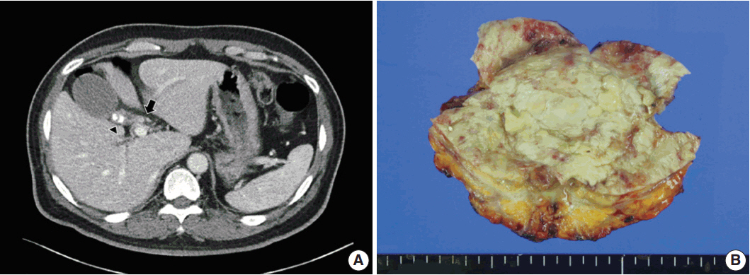
Fig. 1. Computed tomography and gross findings of hyalinizing cholecystitis and associated carcinoma. (A) Biliary computed tomography scan reveals subtle enhancement of wall thickening at the confluence of the cystic duct and the common hepatic duct (arrow). There is mild wall thickening of the gallbladder with multiple calcified gallstones in the neck portion (arrowhead). (B) Grossly, the gallbladder wall shows diffuse fibrosis and is covered by yellowish necrotic materials, without mass-like lesions.
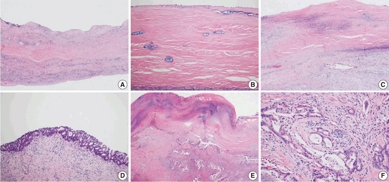
Fig. 2. Microscopic findings of hyalinizing cholecystitis and associated carcinoma. (A, B) The gallbladder wall is replaced by dense lamellated eosinophilic hyaline material. Inflammatory cells are also seen. (C) A few invasive glands are longitudinally arranged in the hyalinized gallbladder wall with a denuded epithelium. (D) Multifocal carcinoma in situ lesions are found on the surface. (E) Focal clusters of invasive glands are identified in the hyalinized wall. (F) Invasive glands have irregular borders and cytologic atypia with perineural invasion.
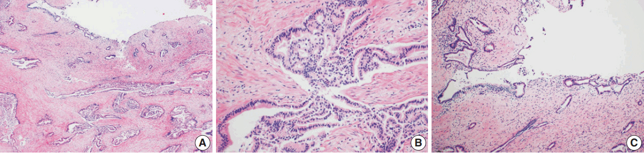
Fig. 3. Microscopic findings of the common hepatic and cystic ducts. (A, B) Invasive glands infiltrate the common hepatic and cystic ducts beneath the surface epithelium. (C) The surface epithelium is relatively spared, with no intraepithelial lesion.
Reference
-
1. Patel S, Roa JC, Tapia O, et al. Hyalinizing cholecystitis and associated carcinomas: clinicopathologic analysis of a distinctive variant of cholecystitis with porcelain-like features and accompanying diagnostically challenging carcinomas. Am J Surg Pathol. 2011; 35:1104–13.2. Kane RA, Jacobs R, Katz J, Costello P. Porcelain gallbladder: ultrasound and CT appearance. Radiology. 1984; 152:137–41.
Article3. Ochsner SF, Carrera GM. Calcification of the gallbladder (“porcelain gallbladder”). Am J Roentgenol Radium Ther Nucl Med. 1963; 89:847–53.4. Weiner PL, Lawson TL. The radiology corner: porcelain gallbladder. Am J Gastroenterol. 1975; 64:224–7.5. Shimizu M, Miura J, Tanaka T, Itoh H, Saitoh Y. Porcelain gallbladder: relation between its type by ultrasound and incidence of cancer. J Clin Gastroenterol. 1989; 11:471–6.6. Stephen AE, Berger DL. Carcinoma in the porcelain gallbladder: a relationship revisited. Surgery. 2001; 129:699–703.
Article7. Towfigh S, McFadden DW, Cortina GR, et al. Porcelain gallbladder is not associated with gallbladder carcinoma. Am Surg. 2001; 67:7–10.8. Gupta RK, Patton KT. Hyalinizing cholecystitis with features of immunoglobulin G4-related disease-coincidence or an unrecognized association? A case report. Hum Pathol. 2015; 46:625–8.
Article
- Full Text Links
-
- Actions
-
Cited
- CITED
-
- Close
- Share
-
- Similar articles
-
- Fine Needle Aspiration Cytology of the Hyalinizing Trabecular Adenoma of the Thyroid Gland: A Case Report
- A Case of Hyalinizing Trabecular Adenoma of the Thyroid Gland
- A Case of Hyalinizing Trabecular Adenoma of the Thyroid Gland
- Cytologic Features of Fine Needle Aspirates of Hyalinizing Trabecular Adenoma with Occult Papillary Carcinoma of the Thyroid
- A Case of Hyalinizing Trabecular Tumor of the Thyroid Gland
