Ann Dermatol.
2018 Feb;30(1):91-93. 10.5021/ad.2018.30.1.91.
A Case of Multiple Cutaneous Piloleiomyomas on the Neck
- Affiliations
-
- 1Department of Dermatology, Soonchunhyang University Seoul Hospital, Seoul, Korea. mkcho2001@hanmail.net
- 2Molecular Cancer Research, Soonchunhyang University College of Medicine, Cheonan, Korea.
- KMID: 2399762
- DOI: http://doi.org/10.5021/ad.2018.30.1.91
Abstract
- No abstract available.
MeSH Terms
Figure
-
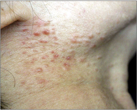
Fig. 1 Multiple erythematous, firm, non-tender papules of various sizes (3~7 mm) on the right side of the neck.

Fig. 2 (A) Dermal proliferation of ill-defined smooth muscle fibers surrounded by varying amounts of collagen fibers (H&E, ×40). (B) Dermal tumor composed of smooth muscle fibers with spindle-shaped, blunt-ended nuclei (H&E, ×400). (C) Tumor cells showing strong positively for smooth muscle actin (smooth muscle actin stain, ×200), and (D) desmin (desmin stain, ×200).
Reference
-
1. Malhotra P, Walia H, Singh A, Ramesh V. Leiomyoma cutis: a clinicopathological series of 37 cases. Indian J Dermatol. 2010; 55:337–341.
Article2. Thyresson HN, Su WP. Familial cutaneous leiomyomatosis. J Am Acad Dermatol. 1981; 4:430–434.
Article3. Menko FH, Maher ER, Schmidt LS, Middelton LA, Aittomäki K, Tomlinson I, et al. Hereditary leiomyomatosis and renal cell cancer (HLRCC): renal cancer risk, surveillance and treatment. Fam Cancer. 2014; 13:637–644.
Article4. Ghanadan A, Abbasi A, Kamyab Hesari K. Cutaneous leiomyoma: novel histologic findings for classification and diagnosis. Acta Med Iran. 2013; 51:19–24.5. Pijpe J, Broers GH, Plaat BE, Hundeiker M, Otto F, Mastik MF, et al. The relation between histological, tumor-biological and clinical parameters in deep and superficial leiomyosarcoma and leiomyoma. Sarcoma. 2002; 6:105–110.
Article
