J Clin Neurol.
2018 Jan;14(1):120-122. 10.3988/jcn.2018.14.1.120.
Ictal Epileptic Headache in an Elderly Patient with a Hippocampal Tumor
- Affiliations
-
- 1Department of Neurology, Haeundae Paik Hospital, Inje University College of Medicine, Busan, Korea. smilepkm@hanmail.net
- KMID: 2399614
- DOI: http://doi.org/10.3988/jcn.2018.14.1.120
Abstract
- No abstract available.
Figure
-
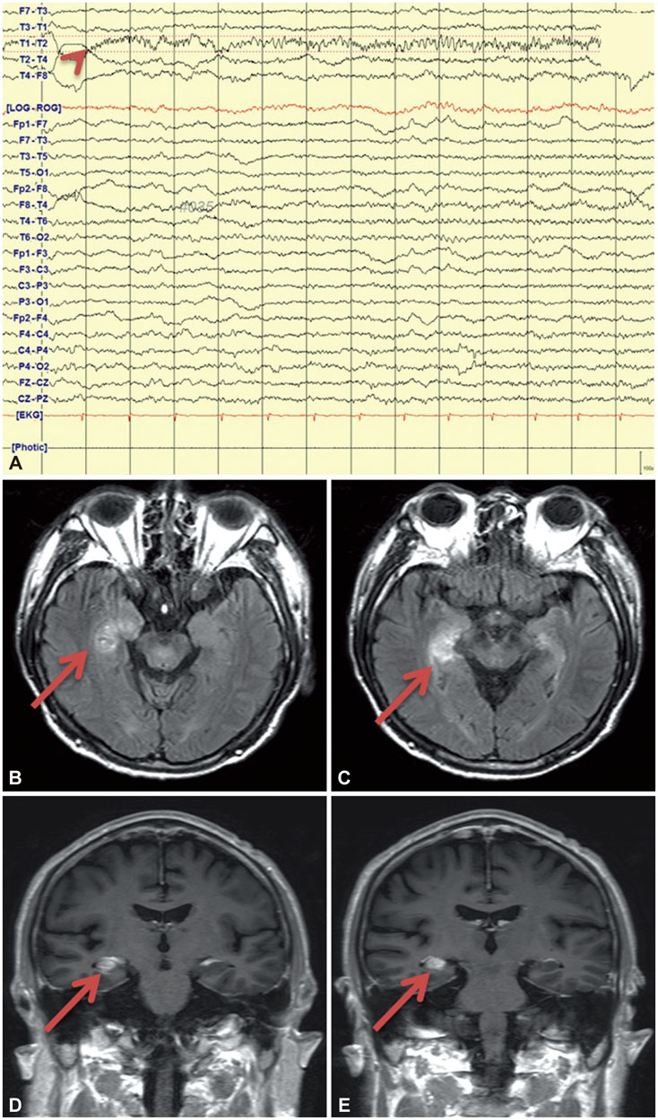
Fig. 1 EEG and Brain MRI. Ictal EEG showing the onset of rhythmic beta activity beginning in the right temporal area (arrowhead) (A). Fluid-attenuated inversion recovery axial (B and C) and T1-weighted coronal (D and E) brain MRI scans showing an enhanced mass lesion in the right hippocampus (arrows).
Reference
-
1. Headache Classification Committee of the International Headache Society (IHS). The International Classification of Headache Disorders, 3rd ed. (beta version). Cephalalgia. 2013; 33:629–808.2. Fanella M, Morano A, Fattouch J, Albini M, Basili LM, Casciato S, et al. Ictal epileptic headache in occipital symptomatic epilepsy: not only a matter of cortex. Headache. 2017; 57:956–961.
Article3. Vydrova R, Kršek P, Kyncl M, Jahodova A, Dvorak J, Komarek V, et al. Peri-ictal headache due to epileptiform activity in a disconnected hemisphere. Epileptic Disord. 2014; 16:213–217.
Article4. Cianchetti C, Pruna D, Porcu L, Peltz MT, Ledda MG. Pure epileptic headache and related manifestations: a video-EEG report and discussion of terminology. Epileptic Disord. 2013; 15:84–92.
Article5. Fanella M, Morano A, Fattouch J, Albini M, Manfredi M, Giallonardo AT, et al. Ictal epileptic headache in adult life: electroclinical patterns and spectrum of related syndromes. Epilepsy Behav. 2015; 53:161–165.
Article
- Full Text Links
-
- Actions
-
Cited
- CITED
-
- Close
- Share
-
- Similar articles
-
- Ictal Cerebral Perfusion Patterns in Partial Epilepsy: SPECT Subtraction
- Ictal Spitting in a Patient with Dominant Temporal Lobe Epilepsy: Discrepancy between Epileptogenic and Symptomatogenic Areas for Spitting Automatism
- Peri-ictal Heart Rate Changes in Patients With Localization-Related Epilepsy
- Incidence and clinical profile of extra-medial-temporal epilepsy with hippocampal atrophy
- The Usefulness of Ictal SPECT in Preoperative Localization of Neocortical Epileptic Foci
