Pyomyoma after Uterine Artery Embolization for Postpartum Hemorrhage Misdiagnosed as Uterine Necrosis
- Affiliations
-
- 1Department of Radiology, Ilsan Paik Hospital, College of Medicine, Inje University, Goyang, Korea.
- 2Department of Radiology, Dongsan Medical Center, Keimyung University School of Medicine, Daegu, Korea. sheen@dsmc.or.kr
- 3Department of Radiology, College of Medicine, Kangwon National University, Chuncheon, Korea.
- KMID: 2399307
- DOI: http://doi.org/10.3348/jksr.2018.78.1.63
Abstract
- A 34-year-old female patient underwent uterine artery embolization (UAE) to control massive postpartum hemorrhage. The interventional radiologist was not informed of the patient's significant history of uterine myoma. Although no significant signs of complications or "red flags" were observed during the procedure, follow-up computed tomography performed four weeks later revealed evidence of a large, globe-like fluid collection with air bubbles in the uterus. The finding and pathology was initially diagnosed as uterine necrosis, which led not to interventional percutaneous drainage; instead, dilation and curettage with resectoscope was performed. The surgical and pathological diagnosis was "expulsion of pyomyoma in the uterine cavity." Awareness and precise knowledge of imaging findings of pyomyoma and uterine necrosis are important for early diagnosis and treatment of UAE-related complications.
MeSH Terms
Figure
-
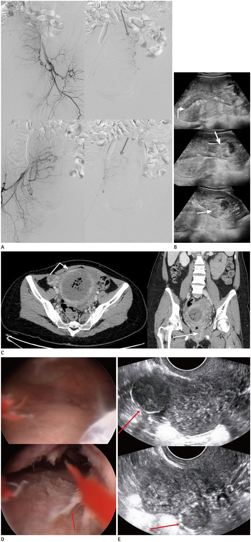
Fig. 1 A 34-year-old female patient with pyomyoma after UAE for postpartum hemorrhage, misdiagnosed as uterine necrosis. A. Angiography of both internal iliac arteries and both uterine arteries was performed on each side. Both uterine arteries (arrows) are engorged, but no active bleeding is detected on angiogram. There is no definitive evidence of myoma on angiography. B. Images of transvaginal uterine ultrasonography 4 days after UAE. Ultrasonography shows hypoechoic area (curved arrow) within uterine cavity, suggesting hematometra. There are also several heterogeneous hyperechoic myomas (arrows) in uterus endometrium and myometrium. UAE = uterine artery embolization C. Images of abdominopelvic computed tomography performed due to fever, abdominal pain and vaginal discharge a month after UAE. Fluid collection with air bubbles and peripheral enhancement in uterine myoma, which is measured about 12.7 × 8.2 × 7.2 cm, is seen in the enlarged uterine cavity. We misdiagnosed this as uterine necrosis, which should have been identified as pyomyoma. Peripheral myometrial enhancement (curved arrow) was diagnostic clue for pyomyoma. In addition, opening of cervix (arrow) by expulsed myoma was diagnostic evidence for pyomyoma. UAE = uterine artery embolization D. Images of hysteroscope performed by obstetrician. Myoma is observed in anterior wall of uterus and suppurative necrotic debris (arrow) was found in uterine cavity on hysteroscope. E. Images of transvaginal uterine ultrasonography nine months after dilation and curettage with resectoscope. Two myomas measuring 2.0 × 1.9 cm and 1.6 × 1.2 cm are noted in myometrium of uterus (arrows), which are measured 4.8 × 2.9 cm and 3.3 × 2.8 cm respectively, on uterine ultrasonography previously performed by obstetrician before UAE. Except for that, there is no remarkable finding in uterus. UAE = uterine artery embolization
Reference
-
1. Courbiere B, Jauffret C, Provansal M, Agostini A, Bartoli JM, Cravello L, et al. Failure of conservative management in postpartum haemorrhage: uterine necrosis and hysterectomy after angiographic selective embolization with gelfoam. Eur J Obstet Gynecol Reprod Biol. 2008; 140:291–293.
Article2. Tseng JJ, Ho JY, Wen MC, Hwang JI. Uterine necrosis associated with acute suppurative myometritis after angiographic selective embolization for refractory postpartum hemorrhage. Am J Obstet Gynecol. 2011; 204:e4–e6.
Article3. Melenhorst M, Hehenkamp W, de Heer K, Ferco HB. CT features in uterine necrosis of unknown cause: a case report. Clin Imaging. 2014; 38:543–546.
Article4. Karcaaltincaba M, Sudakoff GS. CT of a ruptured pyomyoma. AJR Am J Roentgenol. 2003; 181:1375–1377.
Article5. Iwahashi N, Mabuchi Y, Shiro M, Yagi S, Minami S, Ino K. Large uterine pyomyoma in a perimenopausal female: a case report and review of 50 reported cases in the literature. Mol Clin Oncol. 2016; 5:527–531.
Article6. Obele CC, Dunham S, Bennett G, Pagan J, Sung LY, Charles HW. A case of pyomyoma following uterine fibroid embolization and a review of the literature. Case Rep Obstet Gynecol. 2016; 2016:9835412.
Article7. Martins JG, Gaudenti D, Crespo F, Ganesh D, Verma U. Uncommon complication of uterine artery embolization: expulsion of infarcted myoma and uterine sepsis. Case Rep Obstet Gynecol. 2016; 2016:8695318.
Article8. De Vivo A, Mancuso A, Giacobbe A, Savasta LM, De Dominici R, Dugo N, et al. Uterine myomas during pregnancy: a longitudinal sonographic study. Ultrasound Obstet Gynecol. 2011; 37:361–365.
Article9. Poujade O, Ceccaldi PF, Davitian C, Amate P, Chatel P, Khater C, et al. Uterine necrosis following pelvic arterial embolization for post-partum hemorrhage: review of the literature. Eur J Obstet Gynecol Reprod Biol. 2013; 170:309–314.
Article10. Pinto E, Trovão A, Leitão S, Pina C, Mak Fk, Lanhoso A. Conservative laparoscopic approach to a perforated pyomyoma after uterine artery embolization. J Minim Invasive Gynecol. 2012; 19:775–779.
Article
- Full Text Links
-
- Actions
-
Cited
- CITED
-
- Close
- Share
-
- Similar articles
-
- Uterine necrosis after partial obstruction of the uterine artery via selective embolization in postpartum hemorrhage: A case report
- Uterine Necrosis after Uterine Artery Embolization for Postpartum Hemorrhage
- Spontaneous Restoration of Unrecognized Uterine Inversion
- Aberrant Ovarian Artery Originating from the Iliolumbar Artery: A Case Report
- Uterine Artery Embolization as an Effective Management and Diagnostic Tool for Puerperal Uterine Inversion with Severe Postpartum Bleeding: A Case Report
