Glioneuronal Tumor with Neuropil-Like Islands in the Cerebellum: A Case Report
- Affiliations
-
- 1Department of Radiology, Uijeongbu St. Mary's Hospital, College of Medicine, The Catholic University of Korea, Uijeongbu, Korea. violet2@catholic.ac.kr
- 2Department of Pathology, Uijeongbu St. Mary's Hospital, College of Medicine, The Catholic University of Korea, Uijeongbu, Korea.
- 3Department of Neurosurgery, Uijeongbu St. Mary's Hospital, College of Medicine, The Catholic University of Korea, Uijeongbu, Korea.
- KMID: 2399305
- DOI: http://doi.org/10.3348/jksr.2018.78.1.44
Abstract
- Glioneuronal tumor with neuropil-like islands (GTNI) is a rare and novel mixed neuronal-glial tumor that typically affects the supratentorial cerebral hemispheres of adult patients. It is extremely rare for GTNIs to be in the spine of pediatric and adolescent patients, and there have been no reports of infratentorial GTNIs. We report a case of an elderly patient with an anaplastic, infratentorial GTNI that occurred in the cerebellum, including describing MRI features of our case.
MeSH Terms
Figure
-
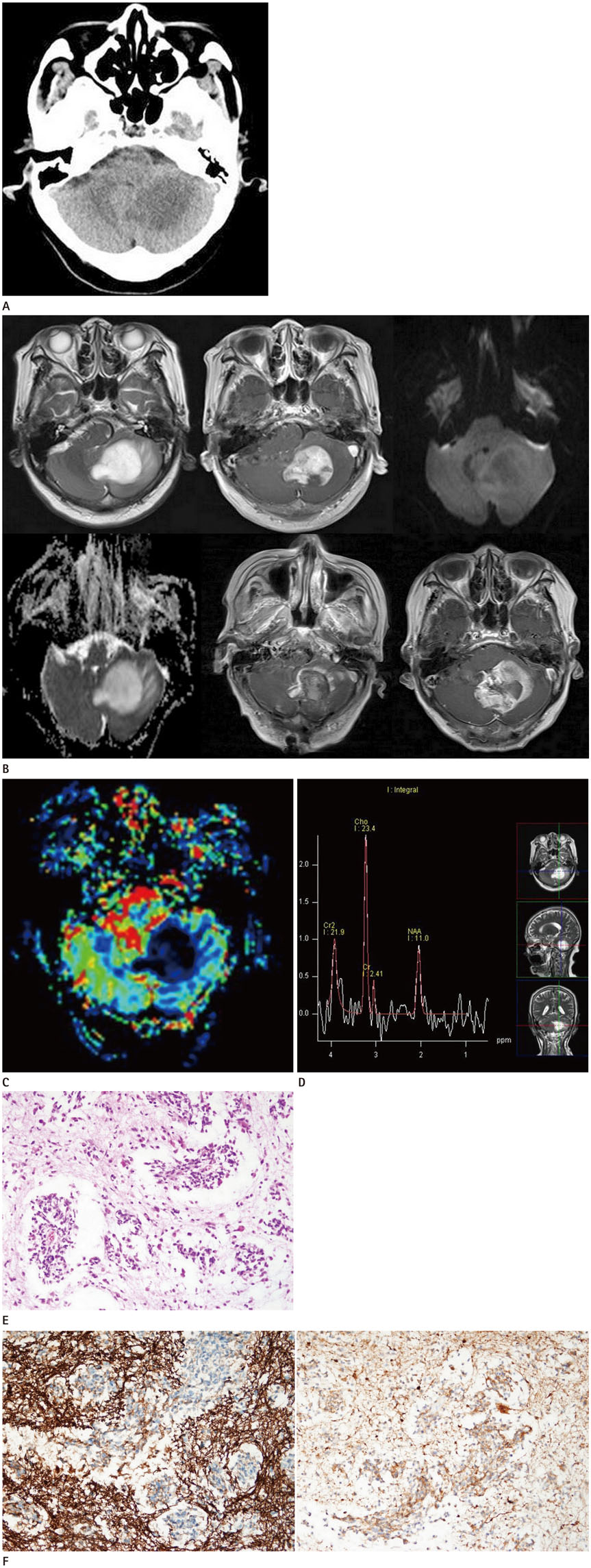
Fig. 1 A 75-year-old woman with anaplastic glioneuronal tumor with neuropil-like islands. A. Unenhanced axial CT scan demonstrates ill-defined hypodensity in the left cerebellar hemisphere. B. The mass exhibits hyper-intensity on axial T2-weighted image (upper left panel) with minimal peritumoral edema and heterogeneously strong enhancement on axial post-contrast T1-weighted image (upper central panel). On diffusion images, the mass exhibits a mainly hypo-intense signal on diffusion weighted image (upper right panel) without diffusion restriction and a higher apparent diffusion coefficient (lower left panel) relative to brain parenchyma. Axial post-contrast T1-weighted image on first day after gross total resection (lower central panel) reveals residual enhancement along the margin of the resection cavity. The three-months postoperative MR image (lower right panel) reveals widespread enhancement with distinct margin in the surgical bed. C. The CBV map reveals low CBV within the lesion. D. Single-voxel intermediate echo time (135 ms) MR spectroscopy demonstrates elevated choline/creatine and choline/N-acetylaspartate peaks. E. Photomicrograph of hematoxylin and eosin stained slide (magnification × 100) reveals spindled or elongated astrocytic cells with well-delineated micronodular, neuropil-like islands and central capillaries. CBV = cerebral blood volume F. Positive glial-fibrillary acid protein stain (left panel, magnification × 100) indicates a glial component. Synaptophysin stain (right panel, magnification × 100) revealed positive staining in part of the tumor, indicating a neuronal component.
Reference
-
1. Louis DN, Ohgaki H, Wiestler OD, Cavenee WK, Burger PC, Jouvet A, et al. The 2007 WHO classification of tumours of the central nervous system. Acta Neuropathol. 2007; 114:97–109.
Article2. Agarwal S, Suri V, Rishi A, Shukla B, Garg A, Sharma MC, et al. Glioneuronal tumor with neuropil-like islands: a new entity. Neuropathology. 2009; 29:96–100.
Article3. Teo JG, Gultekin SH, Bilsky M, Gutin P, Rosenblum MK. A distinctive glioneuronal tumor of the adult cerebrum with neuropil-like (including “rosetted”) islands: report of 4 cases. Am J Surg Pathol. 1999; 23:502–510.4. Park CK, Phi JH, Park SH. Glial tumors with neuronal differentiation. Neurosurg Clin N Am. 2015; 26:117–138.
Article5. Yao K, Duan Z, Li J, Wang Y, Mei X, Li S, et al. Glioneuronal tumor with neuropil-like islands: a histological, immunohistochemical, and molecular study of three cases. Int J Clin Exp Pathol. 2016; 9:7294–7301.6. Poliani PL, Sperli D, Valentini S, Armentano A, Bercich L, Bonetti MF, et al. Spinal glioneuronal tumor with neuropillike islands and meningeal dissemination: histopathological and radiological study of a pediatric case. Neuropathology. 2009; 29:574–578.
Article7. Amemiya S, Shibahara J, Aoki S, Takao H, Ohtomo K. Recently established entities of central nervous system tumors: review of radiological findings. J Comput Assist Tomogr. 2008; 32:279–285.8. Kao HW, Chiang SW, Chung HW, Tsai FY, Chen CY. Advanced MR imaging of gliomas: an update. Biomed Res Int. 2013; 2013:970586.
Article9. Lev MH, Rosen BR. Clinical applications of intracranial perfusion MR imaging. Neuroimaging Clin N Am. 1999; 9:309–331.
- Full Text Links
-
- Actions
-
Cited
- CITED
-
- Close
- Share
-
- Similar articles
-
- Primary Yolk Sac Tumor of the Cerebellar Hemisphere: Case Report
- Dysembryoplastic Neuroepithelial Tumor in the Cerebellum: Case Report
- Immunohistochemical Study on the Distribution of Canonical Transient Receptor Potential Channels in Rat Cerebellum
- A Case of Trichoblastic Fibroma
- Cerebellar Pleomorphic Xanthoastrocytoma: A Case Report
