Arthroscopic detection of medial meniscal injury with the use of a joint distractor in small-breed dogs
- Affiliations
-
- 1College of Veterinary Medicine, Chonbuk National University, Jeonju 54896, Korea.
- 2College of Veterinary Medicine, Chungnam National University, Daejeon 34134, Korea. seatiger76@cnu.ac.kr
- KMID: 2398511
- DOI: http://doi.org/10.4142/jvs.2017.18.4.515
Abstract
- The goal of the present study was to evaluate the diagnostic value of a joint distractor in arthroscopy in small-breed dogs. Sixty stifle joints, which were collected from thirty cadavers, were used in this study. To simulate different injuries, no medial meniscal tear, a full-thickness vertical longitudinal tear, a partial-thickness vertical longitudinal tear, full- and partial-thickness vertical longitudinal tears, or a peripheral detachment were created on the caudal horn of the medial meniscus of each stifle joint along with rupture of the cranial cruciate ligament. Each stifle joint then underwent arthroscopy with and without a joint distractor. The sensitivity (Sn), specificity (Sp), positive predictive value (PPV), negative predictive value (NPV), and correct classification rate (CCR) for the diagnosis of each type of medial meniscus pathology were calculated. For arthroscopy with and without a joint distractor, the Sn was 85% and 60%, the Sp was 96% and 92%, the PPV was 85% and 65%, the NPV was 96% and 90%, and the CCR was 94% and 86%, respectively. Arthroscopy is an effective diagnostic method for the assessment of medial meniscal pathologies in small-breed dogs, especially when performed with the aid of a joint distractor.
Keyword
MeSH Terms
Figure
-

Fig. 1 Schematic diagrams of canine medial meniscal injuries. (A) Normal meniscus. (B) Full-thickness vertical longitudinal tear. (C) Partial-thickness vertical longitudinal tear. (D) Multiple vertical longitudinal tears. (E) Peripheral detachment of the medial meniscus.
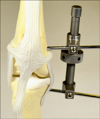
Fig. 2 Photograph of a joint distractor.
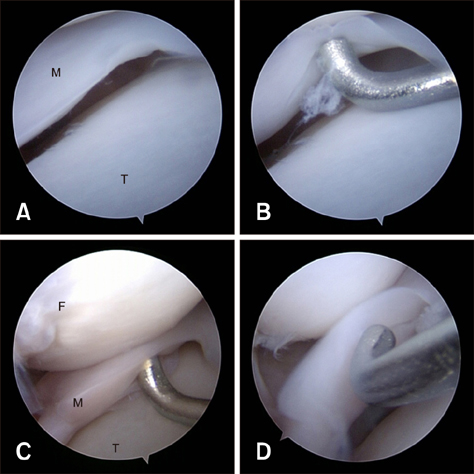
Fig. 3 The arthroscopic appearance of partial-thickness longitudinal medial meniscal tears. (A and B) The same stifle with a joint distractor applied in both cases. (A) An intact meniscus. (B) A tear is revealed by probing. The same stifle without a joint distractor (C) and with a joint distractor (D). By probing, tears were observed in both C and D, but the joint space in C appears narrower than in D. M, meniscus; T, tibia; F, femur.
Reference
-
1. Arnoczky SP, Warren RF. The microvasculature of the meniscus and its response to injury an experimental study in the dog. Am J Sports Med. 1983; 11:131–141.
Article2. Bennett D, May C. Meniscal damage associated with cruciate disease in the dog. J Small Anim Pract. 1991; 32:111–117.
Article3. Böttcher P, Winkels P, Oechtering G. A novel pin distraction device for arthroscopic assessment of the medial meniscus in dogs. Vet Surg. 2009; 38:595–600.
Article4. Bruce W, Rose A, Tuke S, Robins G. Evaluation of the triple tibial osteotomy. A new technique for the management of the canine cruciate-deficient stifle. Vet Comp Orthop Traumatol. 2007; 20:159–168.
Article5. Case J, Hulse D, Kerwin S, Peycke L. Meniscal injury following initial cranial cruciate ligament stabilization surgery in 26 dogs (29 stifles). Vet Comp Orthop Traumatol. 2008; 21:365–367.
Article6. Franklin SP, Gilley RS, Palmer RH. Meniscal injury in dogs with cranial cruciate ligament rupture. Compend Contin Educ Vet. 2010; 32:E1–E10.7. Gemmill TJ, Farrell M. Evaluation of a joint distractor to facilitate arthroscopy of the canine stifle. Vet Surg. 2009; 38:588–594.
Article8. Kim K, Lee HE, Ragetly GR. Feasibility of stifle medial meniscal release in toy breed dogs with and without a joint distractor. Vet Surg. 2016; 45:636–641.
Article9. Metelman LA, Schwarz P, Salman M, Alvis M. An evaluation of three different cranial cruciate ligament surgical stabilization procedures as they relate to postoperative meniscal injuries. Vet Comp Orthop Traumatol. 1995; 8:56–61.
Article10. Nečas A, Zatloukal J. Factors related to the risk of meniscal injury in dogs with cranial cruciate ligament rupture. Acta Vet Brno. 2002; 71:77–84.
Article11. Plesman R, Gilbert P, Campbell J. Detection of meniscal tears by arthroscopy and arthrotomy in dogs with cranial cruciate ligament rupture. A retrospective, cohort study. Vet Comp Orthop Traumatol. 2013; 26:42–46.
Article12. Pozzi A, Hildreth BE, Rajala-Schultz PJ. Comparison of arthroscopy and arthrotomy for diagnosis of medial meniscal pathology: an ex vivo study. Vet Surg. 2008; 37:749–755.
Article13. Ralphs SC, Whitney WO. Arthroscopic evaluation of menisci in dogs with cranial cruciate ligament injuries: 100 cases (1999-2000). J Am Vet Med Assoc. 2002; 221:1601–1604.
Article14. Stein S, Schmoekel H. Short-term and eight to 12 months results of a tibial tuberosity advancement as treatment of canine cranial cruciate ligament damage. J Small Anim Pract. 2008; 49:398–404.
Article15. Steinberg EJ, Prata RG, Palazzini K, Brown DC. Tibial tuberosity advancement for treatment of CrCL injury: complications and owner satisfaction. J Am Anim Hosp Assoc. 2011; 47:250–257.
Article16. Thieman KM, Tomlinson JL, Fox DB, Cook C, Cook JL. Effect of meniscal release on rate of subsequent meniscal tears and ownerXMLLink_XYZassessed outcome in dogs with cruciate disease treated with tibial plateau leveling osteotomy. Vet Surg. 2006; 35:705–710.
Article17. Vasseur PB. Clinical results following nonoperative management for rupture of the cranial cruciate ligament in dogs. Vet Surg. 1984; 13:243–246.
Article
- Full Text Links
-
- Actions
-
Cited
- CITED
-
- Close
- Share
-
- Similar articles
-
- Evaluation of an arthroscopic stifle lever for stifle joint distraction in toy breed dogs
- Avulsion Fracture of The Medial Meniscus: A Case Report
- Comparison of dorsal and medial arthroscopic approach to canine coxofemoral joint: a cadaveric study
- A New Technique of Arthroscopic Meniscal Repair -Modified Inside-Out Technique
- Gait analysis in clinically healthy small to toy breed dogs using a pressure plate
