Outcomes of Arthroscopic Assisted Reduction and Percutaneous Fixation for Tongue-Type Sanders Type II Calcaneal Fractures
- Affiliations
-
- 1Department of Orthopaedic Surgery, Yeungnam University College of Medicine, Daegu, Korea. chpark77@naver.com
- KMID: 2398012
- DOI: http://doi.org/10.14193/jkfas.2017.21.4.144
Abstract
- PURPOSE
To assess the clinical and radiographic results and complications of arthroscopy-assisted reduction and percutaneous fixation for patients with tongue-type Sanders type II calcaneal fractures.
MATERIALS AND METHODS
Between August 2014 and December 2015, 10 patients who underwent surgery using subtalar arthroscopic assisted reduction and percutaneous fixation for tongue-type Sanders type II calcaneal fractures were reviewed. The mean age was 50.8 years (36~62 years), and the mean follow-up period was 24 months (12~40 months). The clinical results were evaluated using the visual analogue scale (VAS) and American Orthopaedic Foot and Ankle Society (AOFAS) ankle-hindfoot score at the regular follow-ups, and the foot function index (FFI) at the last follow-up. The subtalar range of motion (ROM) was evaluated and compared with the uninjured limb at the last follow-up. The radiographic results were assessed using the Böhler's angle from the plain radiographs and the reduction of the posterior calcaneal facet using computed tomography (CT). The postoperative complications were assessed by a chart review.
RESULTS
The VAS and AOFAS ankle-hindfoot score improved until 12 months after surgery. The FFI was 15 (1.8~25.9) and subtalar ROM was 75.5% (60%~100%) compared to the uninjured limb at the last follow-up. The Böhler's angle was increased significantly from 2° (−14°~18°) preoperatively to 21.8° (20°~28°) at the last follow-up. The reduction of the posterior facet was graded as excellent in five feet (50.0%) and good in five (50.0%) on CT obtained at 12 months after surgery. One foot (10.0%) had subfibular pain due to a prominent screw head. One foot (10.0%) had pain due to a longitudinal tear of the peroneal tendon that occurred during screw insertion.
CONCLUSION
Subtalar arthroscopic-assisted reduction of the posterior calcaneal facet of the subtalar joint and percutaneous fixation is a useful surgical method for tongue-type Sanders type II calcaneal fractures.
MeSH Terms
Figure
-
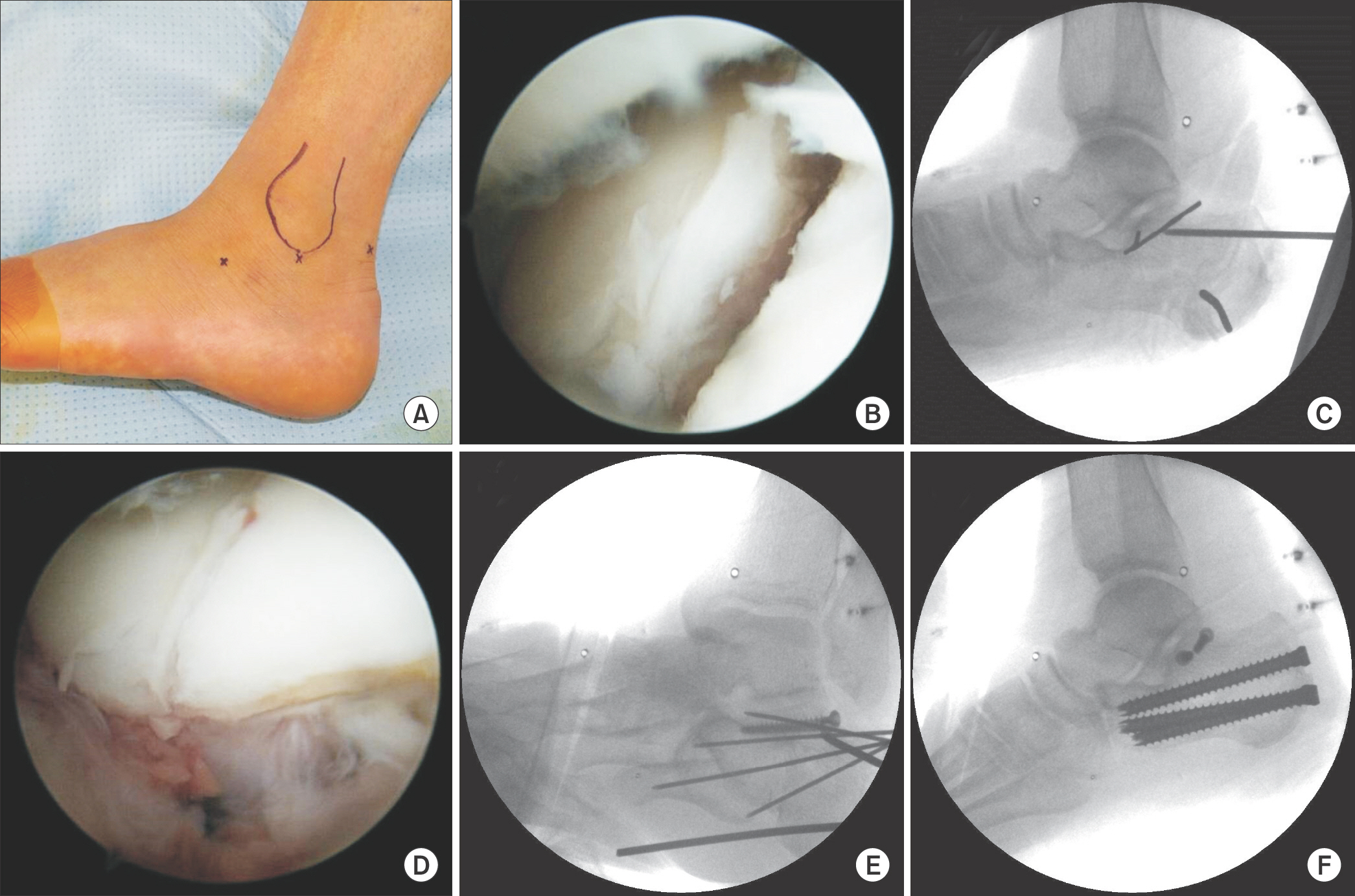
Figure 1. (A) Three subtalar arthroscopic portals (anterolateral, centrolateral, and posterolateral). The anterolateral portal is made 1 cm distal and 2 cm anterior to the tip of the lateral malleolus. The centrolateral portal is made just anterior to the tip of the lateral malleolus. (B) Fracture site of the calcaneus is visualized through anterolateral portal after debridement of hematoma using shaver. (C) Steinmann pin of 2.4 mm is inserted at the tongue-type fragment for the leverage technique. (D) Reduction of the posterior facet is achieved using leverage technique. (E) Medial and lateral fragments of calcaneus are fixated using two 3.5 mm cortical screws. (F) Anterior and posterior fragments are fixated using two or three 7.0 mm cannulated screws.
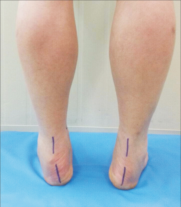
Figure 2. Subtalar range of motion are assessed by measuring the angle between Achilles tendon and calcaneus in full inversion position.
Cited by 1 articles
-
Surgical Treatment of Calcaneal Fractures of Sanders Type II and III by A Minimally Invasive Technique with 6.5 mm Cancellous Screw
Yong Seung Oh, Kyung Ho Lee, Jung Ho Kim, Myoung Jin Lee
J Korean Foot Ankle Soc. 2019;23(3):116-120. doi: 10.14193/jkfas.2019.23.3.116.
Reference
-
11.Epstein N., Chandran S., Chou L. Current concepts review: intra-articular fractures of the calcaneus1 Foot Ankle Int1. 2012. 33:79–861.21.Sanders R. Displaced intra-articular fractures of the calcaneus1 J Bone Joint Surg Am1. 2000. 82:225–501.31.Buckley R., Tough S., McCormack R., Pate G., Leighton R., Petrie D, et al. Operative compared with nonoperative treatment of displaced intra-articular calcaneal fractures: a prospective, randomized, controlled multicenter trial1 J Bone Joint Surg Am1. 2002. 84:1733–441.41.Buckley RE., Meek RN. Comparison of open versus closed reduction of intra-articular calcaneal fractures: a matched cohort in workmen1 J Orthop Trauma1. 1992. 6:195–2051.51.Sanders R., Fortin P., DiPasquale T., Walling A. Operative treatment in 120 displaced intraarticular calcaneal fractures1 Results using a prognostic computed tomography scan classification1 Clin Orthop Relat Res1. 1993. 290:87–951.61.Kim WY., Ji JH., Kwon OS., Park SE., Kim YY., Kil HJ, et al. Clinical features of distal tibial fractures and treatment results of minimally invasive plate osteosynthesis1 J Korean Foot Ankle Soc1. 2012. 16:94–1001.71.Kim B., Lee G., Jang H. Minimal invasive plate osteosynthesis versus conventional open plating in simple humeral shaft fracture (AO Type A, B1, B2)1 J Korean Fract Soc1. 2017. 30:124–301.81.Zwipp H., Rammelt S., Barthel S. Calcaneal fractures--open reduction and internal fixation (ORIF)1 Injury1. 2004. 35(Suppl 2):SB46-541.91.Freeman BJ., Duff S., Allen PE., Nicholson HD., Atkins RM. The extended lateral approach to the hindfoot1 Anatomical basis and surgical implications1 J Bone Joint Surg Br1. 1998. 80:139–421.101.DeWall M., Henderson CE., McKinley TO., Phelps T., Dolan L., Marsh JL. Percutaneous reduction and fixation of displaced intra-articular calcaneus fractures1 J Orthop Trauma1. 2010. 24:466–721.111.Pastides PS., Milnes L., Rosenfeld PF. Percutaneous arthroscopic calcaneal osteosynthesis: a minimally invasive technique for displaced intra-articular calcaneal fractures1 J Foot Ankle Surg1. 2015. 54:798–8041.121.Park CH., Lee DY. Surgical treatment of sanders type 2 calcaneal fractures using a sinus tarsi approach1 Indian J Orthop1. 2017. 51:461–71.131.Westhues H. Eine neue behadungsmethode der calcaneus frakturen1 Zbl Chir. 1935. 17:995–10021.141.Gissane W. Discussion on “Fractures of the os calcis”1 Proceedings of the British Orthopaedic Association1 J Bone Joint Surg Am1. 1947. 29:254–51.151.Essex-Lopresti P. The mechanism, reduction technique, and results in fractures of the os calcis1 Br J Surg1. 1952. 39:395–4191.161.Rammelt S., Gavlik JM., Barthel S., Zwipp H. The value of subtalar arthroscopy in the management of intra-articular calcaneus fractures1 Foot Ankle Int1. 2002. 23:906–161.171.Woon CY., Chong KW., Yeo W., Eng-Meng Yeo N., Wong MK. Subtalar arthroscopy and flurosocopy in percutaneous fixation of intra-articular calcaneal fractures: the best of both worlds1 J Trauma1. 2011. 71:917–251.181.Chung KC., Gwak HC., Kim CW., Kim JH., Park DH. The usefulness of arthroscopy in the operative treatment of intra-articular calcaneal fracture1 J Korean Foot Ankle Soc1. 2009. 13:55–91.191.Rammelt S., Amlang M., Barthel S., Gavlik JM., Zwipp H. Percutaneous treatment of less severe intraarticular calcaneal fractures1 Clin Orthop Relat Res1. 2010. 468:983–901.201.Park CH., Kim SY., Kim JR., Lee WC. Arthroscopic excision of a symptomatic os trigonum in a lateral decubitus position1 Foot Ankle Int1. 2013. 34:990–41.211.Kurozumi T., Jinno Y., Sato T., Inoue H., Aitani T., Okuda K. Open reduction for intra-articular calcaneal fractures: evaluation using computed tomography1 Foot Ankle Int1. 2003. 24:942–81.221.Sivakumar BS., Wong P., Dick CG., Steer RA., Tetsworth K. Ar-throscopic reduction and percutaneous fixation of selected calcaneus fractures: surgical technique and early results1 J Orthop Trauma1. 2014. 28:569–761.231.Benirschke SK., Kramer PA. Wound healing complications in closed and open calcaneal fractures1 J Orthop Trauma1. 2004. 18:1–61.241.Levin LS., Nunley JA. The management of soft-tissue problems associated with calcaneal fractures1 Clin Orthop Relat Res1. 1993. 290:151–61.251.Howard JL., Buckley R., McCormack R., Pate G., Leighton R., Petrie D, et al. Complications following management of displaced intra-articular calcaneal fractures: a prospective randomized trial comparing open reduction internal fixation with nonoperative management1 J Orthop Trauma1. 2003. 17:241–91.261.Rammelt S., Amlang M., Barthel S., Zwipp H. Minimally-invasive treatment of calcaneal fractures1 Injury1. 2004. 35(Suppl 2):SB55-631.271.Tornetta P 3rd. Percutaneous treatment of calcaneal fractures1 Clin Orthop Relat Res1. 2000. 375:91–61.281.Broden B. Roentgen examination of the subtaloid joint in fractures of the calcaneus1 Acta radiol1. 1949. 31:85–911.291.Gavlik JM., Rammelt S., Zwipp H. Percutaneous, arthroscopically-assisted osteosynthesis of calcaneus fractures1 Arch Orthop Trauma Surg1. 2002. 122:424–81.301.Chung SW., Park JS., Kim SH., Shin SH., Oh JH. Quality of life after arthroscopic rotator cuff repair: evaluation using SF-36 and an analysis of affecting clinical factors1 Am J Sports Med1. 2012. 40:631–91.
- Full Text Links
-
- Actions
-
Cited
- CITED
-
- Close
- Share
-
- Similar articles
-
- The Usefulness of Arthroscopy in the Operative Treatment of Intra-Articular Calcaneal Fracture
- Minimally-invasive Percutaneous Screw Fixation of Displaced Intra-articular Calcaneal Fractures
- Treatment of Intra-articular Calcaneal Fracture with Open Reduction and Internal Fixation
- Surgical Outcomes of Intra-articular Fractures of Calcaneus using AO Calcaneal Plate
- Usefulness of CT Scan in Treatment of Calcaneal Fracture
