Korean Circ J.
2017 Nov;47(6):978-980. 10.4070/kcj.2017.0115.
Huge Fresh Mobile Thrombus Attached to the Descending Thoracic Aorta
- Affiliations
-
- 1Department of Cardiology in Internal Medicine, Chungnam National University School of Medicine, Chungnam National University Hospital, Daejeon, Korea. jaehpark@cnuh.co.kr
- KMID: 2396491
- DOI: http://doi.org/10.4070/kcj.2017.0115
Abstract
- No abstract available.
MeSH Terms
Figure
-
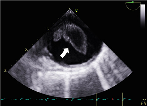
Figure 1 On the transesophageal echocardiographic examination, a 1.5×0.7 cm sized, highly mobile mass lesion is found in the descending thoracic aorta (arrow).

Figure 2 A linear, non-enhanced, low-attenuated mass lesion suggesting a thrombus is found at the distal aortic arch on contrast-enhanced spiral aorta computerized tomography scan (arrow, A: axial view, B: coronal view).
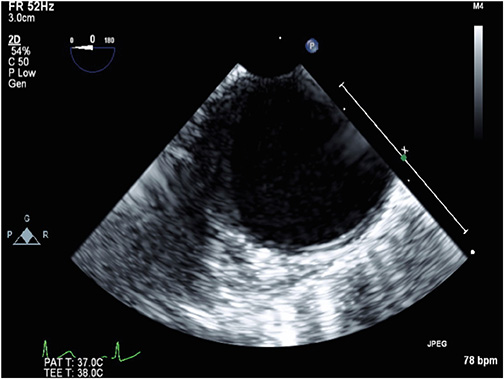
Figure 3 The huge free-floating mass disappeared on the follow-up transesophageal echocardiographic examination after 2 weeks of anticoagulation therapy.
Reference
-
1. Dee W, Geibel A, Kasper W, Konstantinides S, Just H. Mobile thrombi in atherosclerotic lesions of the thoracic aorta: the diagnostic impact of transesophageal echocardiography. Am Heart J. 1993; 126:707–710.2. Laperche T, Laurian C, Roudaut R, Steg PG. Mobile thromboses of the aortic arch without aortic debris. A transesophageal echocardiographic finding associated with unexplained arterial embolism. The Filiale Echocardiographie de la Société Française de Cardiologie. Circulation. 1997; 96:288–294.3. French Study of Aortic Plaques in Stroke Group. Amarenco P, Cohen A, et al. Atherosclerotic disease of the aortic arch as a risk factor for recurrent ischemic stroke. N Engl J Med. 1996; 334:1216–1221.
- Full Text Links
-
- Actions
-
Cited
- CITED
-
- Close
- Share
-
- Similar articles
-
- Floating Thrombus in Aortic Arch
- Acute Abdominal Mobile Aortic Thrombus Post Chemotherapy: Two Cases Reports
- Cerebral Infarction Caused by Floating Thoracic aortic Thrombus in Young Male
- A Case of Mobile Thrombus in Ascending Aorta as an Embolic Source of Stroke
- Successful hybrid operation of an acute mobile thrombus in the abdominal aorta induced by chemotherapy
