Checkrein Deformity by Incarcerated Posterior Tibial Tendon and Displaced Flexor Hallucis Longus Tendon following Ankle Dislocation: A Case Report
- Affiliations
-
- 1Department of Orthopaedic Surgery, Sanggye Paik Hospital, Inje University, Seoul, Korea. sybae99@gmail.com
Abstract
- We report a case of 20 year-old man who had unusual equinus and checkrein deformity following dislocation of his right ankle joint. He had been treated with distal tibiofibular screw fixation and external fixation. After removal of external fixator, he had suffered from progressive deformity of foot and ankle. Widening of distal tibiofibular joint and medial clear space was found on radiograph and it was revealed that posterior tibial tendon had been dislocated and incarcerated into the distal tibiofibular joint on MRI. We corrected the deformity with excision of incarcerated posterior tibial tendon, adhesiolysis and lengthening of flexor hallucis longus tendon, reconstruction of deltoid ligament and flexor digitorum longus tendon transfer.
MeSH Terms
Figure
-
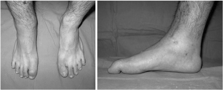
Fig. 1 Equinus of right ankle and flexion deformity of interphalangeal joint of great toe.
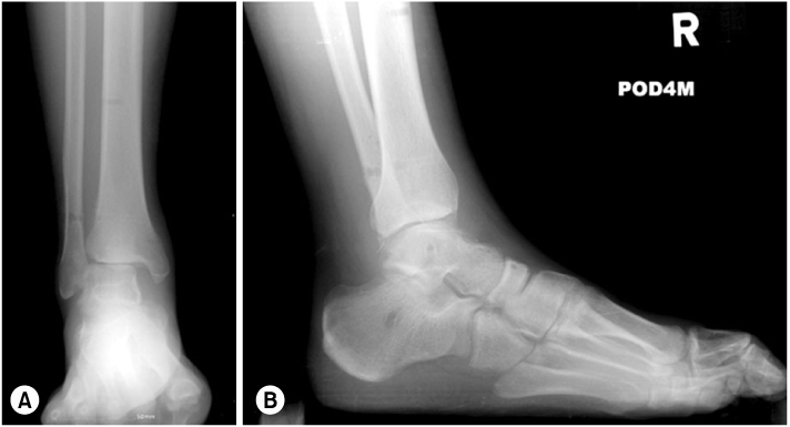
Fig. 2 (A) Anteroposterior view of right ankle shows widening of tibiofibular space and medial clear space. (B) Lateral view shows equinus and flexion deformity of great toe.
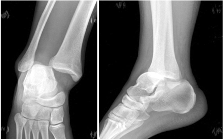
Fig. 3 Immediate post-traumatic radiographs of other clinic show that talus was displaced into distal tibiofibular space.
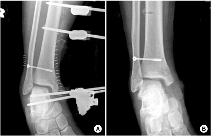
Fig. 4 (A) For initial treatment, open reduction and external fixation of ankle joint was done and a syndesmotic screw was inserted at other clinic. (B) Follow-up X-ray with the external fixator removed 2 months later.

Fig. 5 Ankle MRI shows incarceration of posterior tibial tendon into the fibular groove of distal tibia (white arrows) and displaced flexor hallucis longus tendon (black arrows).
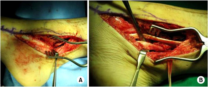
Fig. 6 (A) Posterior tibial tendon was displaced into the posterolateral aspect of the ankle joint. (B) Continuity of flexor hallucis longus tendon was intact (freer under the tendon).
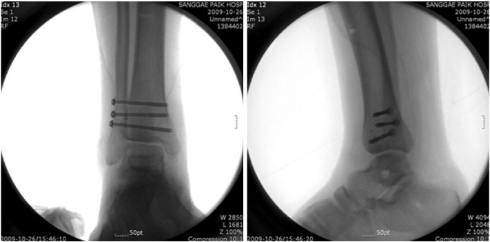
Fig. 7 Intra-operative ankle X-ray (AP and lateral views).
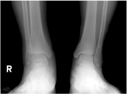
Fig. 8 Follow-up standing ankle radiograph.
Reference
-
1. Carr JB. Complication of calcaneus fractures entrapment of the flexor hallucis longus: report of two cases. J Orthop Trauma. 1990. 4:166–168.
Article2. De Zwart DF, Davidson JS. Rupture of the posterior tibial tendon associated with fractures of the ankle. A report of two cases. J Bone Joint Surg Am. 1983. 65:260–262.
Article3. Ermis MN, Yagmurlu MF, Kilinc AS, Karakas ES. Irreducible fracture dislocation of the ankle caused by tibialis posterior tendon interposition. J Foot Ankle Surg. 2010. 49:166–171.
Article4. Feeney MS, Williams RL, Stephens MM. Selective lengthening of the proximal flexor tendon in the management of acquired claw toes. J Bone Joint Surg Br. 2001. 83:335–338.
Article5. Jahss MH. Disorders of the foot and ankle. 1991. Vol. 2:2nd ed. Philadelphia: WB Saunders;1471–1477.6. Kwon H, Kim DW, Kim DJ, Sohn CS, Song JM, Rah SK. Checkrein deformity of the lesser toes following comminuted fracture of calcaneus: a case report. J Korean Soc Fract. 1998. 11:806–810.
Article7. Lee HS, Kim JS, Park SS, Lee DH, Park JM, Wapner KL. Treatment of checkrein deformity of the hallux. J Bone Joint Surg Br. 2008. 90:1055–1058.
Article8. Myerson M, Corrigan J. Treatment of posterior tibialis tendon dysfunction with flexor digitorum longus tendon transfer and calcaneal osteotomy. Orthopedics. 1996. 19:383–388.
Article9. Sanhudo JA, Lompa PA. Checkrein deformity--flexor hallucis tethering: two case reports. Foot Ankle Int. 2002. 23:799–800.
Article
- Full Text Links
-
- Actions
-
Cited
- CITED
-
- Close
- Share
-
- Similar articles
-
- Checkrein Deformity Due to Flexor Digitorum Longus Adhesion after Comminuted Calcaneus Fracture: A Case Report
- The Checkrein Deformity of Extensor Hallucis Longus Tendon and Extensor Retinaculum Syndrome with Deep Peroneal Nerve Entrapment after Triplane Fracture: A Case Report
- Management of Checkrein Deformity
- Proximal Flexor Hallucis Longus Tendon Transfer for the Ossification of the Achilles Tendon: A Case Report
- Checkrein deformity of the lesser toes following comminuted fracture of calcaneus : A Case Report
