Yonsei Med J.
2008 Feb;49(1):111-118.
Imaging Findings of Implanted Absorbable Mesh in Patients with Breast Partial Resection
Abstract
- PURPOSE
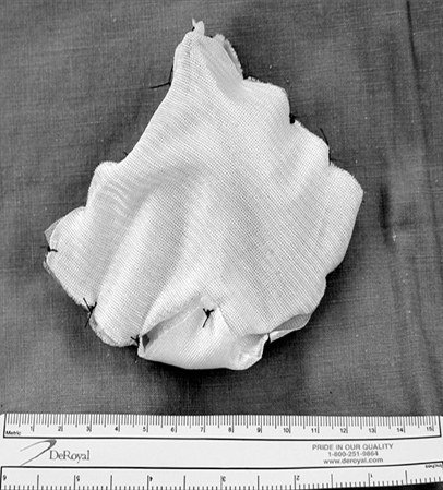
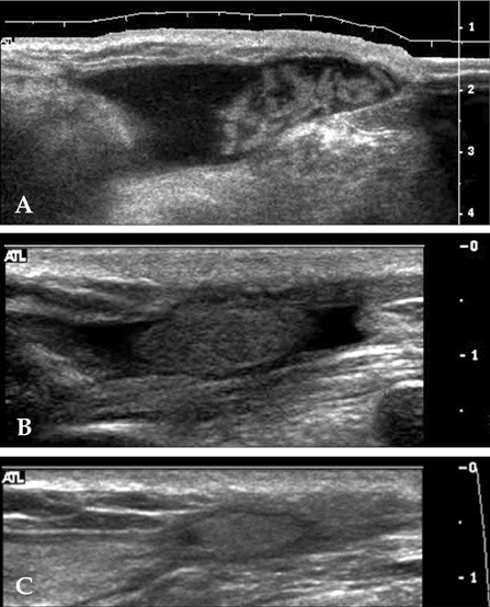
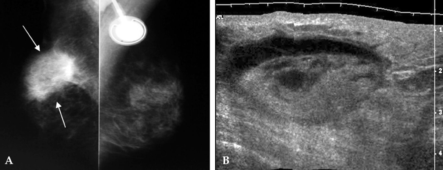
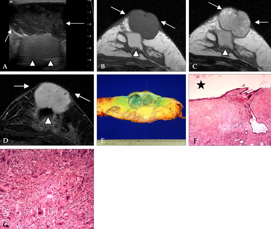
The author presents imaging findings of patients that underwent partial resection of the breast followed by absorbable mesh implantation. MATERIALS AND METHODS: Ultrasonographic (n=18) and mammographic (n=11) images of patients that had undergone absorbable mesh implantation after breast partial resection were reviewed retrospectively. Sequential changes of the lesions were analyzed in follow-up ultrasonographic examinations, focusing on the change of the size and pattern of the lesion. The presence of a mass, asymmetry, focal asymmetry, architectural distortion, and calcification were evaluated by mammography. Pathologic findings of the implanted mesh in available cases were analyzed. RESULTS: Ultrasonograms revealed a well-encapsulated anechoic lesion with (pattern 1, n=11) or without (pattern 2, n=5) internal isoechoic nodular portion, and a hyperechoic mass-like lesion without anechoic portion (pattern 3, n=2). The mean length of the longest diameter decreased gradually as determined in follow-up examinations (3 months, 6.12 +/- 2.599cm; 6 months, 5.08 +/- 2.105cm; 12 months, 3.26 +/- 2.206cm). In mammograms, a mass (n=4) was noted at the surgical site and focal asymmetry, overlapping with the postoperative change, was seen in the remaining seven cases. Pathologic findings of two cases revealed foreign body reaction. CONCLUSION: Ultrasonography of the patients that underwent breast partial resection followed by absorbable mesh implantation showed a well-encapsulated cyst at the surgical site that gradually decreased in follow-up examinations. Adjunctive ultrasonography combined with mammography would be recommended in postoperative follow-up examinations.
Keyword
MeSH Terms
Figure
Reference
-
1. Veronesi U, Cascinelli N, Mariani L, Greco M, Saccozzi R, Luini A, et al. Twenty-year follow-up of a randomized study comparing breast-conserving surgery with radical mastectomy for early breast cancer. N Engl J Med. 2002. 347:1227–1232.
Article2. Choi Y, Hong HP, Kwag HJ. Ultrasonographic findings of an implanted absorbable mesh in patients with breast partial resection: a preliminary study. J Korean Soc Ultrasound Med. 2007. 26:89–94.3. Tobias AM, Low DW. The use of a subfascial vicryl mesh buttress to aid in the closure of massive ventral hernias following damage-control laparotomy. Plast Reconstr Surg. 2003. 112:766–776.
Article4. Dayton MT, Buchele BA, Shirazi SS, Hunt LB. Use of an absorbable mesh to repair contaminated abdominalwall defects. Arch Surg. 1986. 121:954–960.
Article5. Wainstein MA, Resnick MI. Use of polyglycolic acid mesh to support parenchymal closure following partial nephrectomy. J Urol. 1997. 158:526–527.
Article6. Rogers FB, Baumgartner NE, Robin AP, Barrett JA. Absorbable mesh splenorrhaphy for severe splenic injuries: functional studies in an animal model and an additional patient series. J Trauma. 1991. 31:200–204.7. Berry MF, Rosato EF, Williams NN. Dexon mesh splenorrhaphy for intraoperative splenic injuries. Am Surg. 2003. 69:176–180.8. Goes JC. Periareolar mammaplasty: double skin technique with application of polyglactine or mixed mesh. Plast Reconstr Surg. 1996. 97:959–968.
Article9. Sanuki J, Fukuma E, Wadamori K, Higa K, Sakamoto N, Tsunoda Y. Volume replacement with polyglycolic acid mesh for correcting breast deformity after endoscopic conservative surgery. Clin Breast Cancer. 2005. 6:175.
Article10. Boulanger L, Boukerrou M, Lambaudie E, Defossez A, Cosson M. Tissue integration and tolerance to meshes used in gynecologic surgery: an experimental study. Eur J Obstet Gynecol Reprod Biol. 2006. 125:103–108.
Article11. Krause HG, Galloway SJ, Khoo SK, Lourie R, Goh JT. Biocompatible properties of surgical mesh using an animal model. Aust N Z J Obstet Gynaecol. 2006. 46:42–45.
Article12. Klinge U, Schumpelick V, Klosterhalfen B. Functional assessment and tissue response of short- and long-term absorbable surgical meshes. Biomaterials. 2001. 22:1415–1424.
Article13. Barbolt TA. Biology of polypropylene/polyglactin 910 grafts. Int Urogynecol J Pelvic Floor Dysfunct. 2006. 17:Suppl 1. S26–S30.
Article14. Mounzer AM, McAninch JW, Schmidt RA. Polyglycolic acid mesh in repair of renal injury. Urology. 1986. 28:127–130.
Article15. Orel SG, Troupin RH, Patterson EA, Fowble BL. Breast cancer recurrence after lumpectomy and irradiation: role of mammography in detection. Radiology. 1992. 183:201–206.
Article16. Grosse A, Schreer I, Frischbier HJ, Maass H, Loening T, Bahnsen J. Results of breast conserving therapy for early breast cancer and the role of mammographic follow-up. Int J Radiat Oncol Biol Phys. 1997. 38:761–767.
Article17. Hassell PR, Olivotto IA, Mueller HA, Kingston GW, Basco VE. Early breast cancer: detection of recurrence after conservative surgery and radiation therapy. Radiology. 1990. 176:731–735.
Article18. Dershaw DD, McCormick B, Osborne MP. Detection of local recurrence after conservative therapy for breast carcinoma. Cancer. 1992. 70:493–496.
Article19. Giess CS, Keating DM, Osborne MP, Rosenblatt R. Local tumor recurrence following breast-conservation therapy: correlation of histopathologic findings with detection method and mammographic findings. Radiology. 1999. 212:829–835.
Article20. Liberman L, Van Zee KJ, Dershaw DD, Morris EA, Abramson AF, Samli B. Mammographic features of local recurrence in women who have undergone breast-conserving therapy for ductal carcinoma in situ. Am J Roentgenol. 1997. 168:489–493.
Article21. Philpotts LE, Lee CH, Haffty BG, Lange RC, Tocino I. Mammographic findings of recurrent breast cancer after lumpectomy and radiation therapy: comparison with the primary tumor. Radiology. 1996. 201:767–771.
Article22. Goes JC, Landecker A, Lyra EC, Henriquez LJ, Goes RS, Godoy PM. The application of mesh support in periareolar breast surgery: clinical and mammographic evaluation. Aesthetic Plast Surg. 2004. 28:268–274.
Article
- Full Text Links
- Actions
-
Cited
- CITED
-
- Close
- Share
- Similar articles
-
- Ultrasonographic Findings of an Implanted Absorbable Mesh in Patients with Breast Partial Resection: a Preliminary Study
- The Use of Absorbable Surgical Mesh after Partial Mastectomy for Improving the Cosmetic Outcome
- The Suitability of Absorbable Mesh Insertion for Oncoplastic Breast Surgery in Patients with Breast Cancer Scheduled to Be Irradiated
- Nationwide Survey of the Use of Absorbable Mesh in Breast Surgery in Korea
- Results of Absorbable Mesh Insertion and Patient Satisfaction in Breast-Conserving Surgery


