Yonsei Med J.
2009 Apr;50(2):206-210.
Inaccuracy of Intraocular Lens Power Prediction for Cataract Surgery in Angle-Closure Glaucoma
- Affiliations
-
- 1Institute of Vision Research, Department of Ophthalmology, Yonsei University College of Medicine, Seoul, Korea. kcyeye@yuhs.ac
Abstract
- PURPOSE
To assess the accuracy of intraocular lens (IOL) power predictions for cataract surgery in eyes with primary angle-closure glaucoma (ACG). Because of shifting of the capsular bag apparatus and shortening of the axial length, preoperative calculation of IOL power may be inaccurate for eyes with ACG. MATERIALS AND METHODS: This retrospective comparative case series comprised of 42 eyes from 42 patients with primary ACG and 45 eyes from 45 subjects with normal open-angles undergoing uneventful cataract surgery. Anterior segment biometry including anterior chamber depth, lens thickness, and axial length were compared. Using the SRK-II formula, the powers of the implanted IOL and the actual postoperative spherical equivalent (SE) refractive errors were compared between the two groups. Also, the absolute values of differences between predicted and residual SE refractive errors were also analyzed for each group. RESULTS: In ACG patients, anterior chamber depth and axial length were shorter and the lens was thicker than normal controls (all p < 0.001). Even though residual SE refractive error was not significantly different (p = 0.290), the absolute value of the difference between predicted and residual SE refractive error was 0.64 +/- 0.50 diopters in AGC patients and 0.39 +/- 0.36 diopters in control subjects (p = 0.012). The number of eyes that resulted in inaccurate IOL power predictions of more than 0.5 diopters were 21 (50.00%) in the ACG group, but only 12 (26.67%) in the control group (p = 0.043). CONCLUSION: IOL power predictions for cataract surgery in ACG patients can be inaccurate, and it may be associated with their unique anterior segment anatomy.
Keyword
MeSH Terms
Figure
-

Fig. 1 Possible anatomic changes after cataract surgery in eyes with angle closure glaucoma. (A) Hyperopic shift can be caused by deepening of the anterior chamber and shortening of the axial length. (B) Myopic and/or hyperopic shift by tilted or decentered intraocular lens due to the large capsular bag.
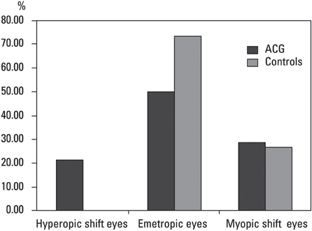
Fig. 2 Subgroups according to the accuracy of the intraocular lens power prediction after cataract surgery in angle closure glaucoma patients and normal controls. ACG, angle-closure glaucoma.
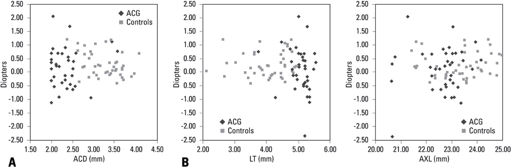
Fig. 3 Correlation between anterior segment biometry and accuracy of intraocular lens power calculation. (A) Anterior chamber depth (ACD), (B) Lens thickness (LT), (C) Axial length (AXL) in eyes with angle closure glucoma.
Reference
-
1. Yang CH, Hung PT. Intraocular lens position and anterior chamber angle changes after cataract extraction in eyes with primary angle-closure glaucoma. J Cataract Refract Surg. 1997. 23:1109–1113.
Article2. Hayashi K, Hayashi H, Nakao F, Hayashi F. Changes in anterior chamber angle width and depth after intraocular lens implantation in eyes with glaucoma. Ophthalmology. 2000. 107:698–703.
Article3. Nonaka A, Kondo T, Kikuchi M, Yamashiro K, Fujihara M, Iwawaki T, et al. Angle widening and alteration of ciliary process configuration after cataract surgery for primary angle closure. Ophthalmology. 2006. 113:437–441.
Article4. Francis BA, Wang M, Lei H, Du LT, Minckler DS, Green RL, et al. Changes in axial length following trabeculectomy and glaucoma drainage device surgery. Br J Ophthalmol. 2005. 89:17–20.
Article5. Law SK, Mansury AM, Vasudev D, Caprioli J. Effects of combined cataract surgery and trabeculectomy with mitomycin C on ocular dimensions. Br J Ophthalmol. 2005. 89:1021–1025.
Article6. Lowe RF. Aetiology of the anatomical basis for primary angle-closure glaucoma. Biometrical comparisons between normal eyes and eyes with primary angle-closure glaucoma. Br J Ophthalmol. 1970. 54:161–169.
Article7. Markowitz SN, Morin JD. Angle-closure glaucoma: relation between lens thickness, anterior chamber depth and age. Can J Ophthalmol. 1984. 19:300–302.8. Marchini G, Pagliarusco A, Toscano A, Tosi R, Brunelli C, Bonomi L. Ultrasound biomicroscopic and conventional ultrasonographic study of ocular dimensions in primary angle-closure glaucoma. Ophthalmology. 1998. 105:2091–2098.
Article
- Full Text Links
-
- Actions
-
Cited
- CITED
-
- Close
- Share
-
- Similar articles
-
- Clinical Factors that Influence Intraocular Pressure Change after Cataract Surgery in Primary Open-Angle Glaucoma and Angle-Closure Glaucoma
- IOP Change after Cataract Surgery in Patients with Controlled Chronic Glaucoma
- The Change of Intraocular Pressure after Extracapsular Cataract Extraction in Patients with Angle-closure Glaucoma
- The Combined Cataract Extraction and Cyclodialysis for the Secondary Angle-Closure Glaucoma Due to the Intumescent Cataract
- Iris-trabecular Contact Index Change after Cataract Surgery in Acute Angle Closure Glaucoma
