Ann Dermatol.
2017 Apr;29(2):167-172. 10.5021/ad.2017.29.2.167.
Rosacea Subtypes Visually and Optically Distinct When Viewed with Parallel-Polarized Imaging Technique
- Affiliations
-
- 1Department of Dermatology, Korea University College of Medicine, Seoul, Korea. gold2000@nate.com
- KMID: 2394839
- DOI: http://doi.org/10.5021/ad.2017.29.2.167
Abstract
- BACKGROUND
Parallel-polarized light (PPL) photography evaluates skin characteristics by analyzing light reflections from the skin surface.
OBJECTIVE
The aim of this study was to determine the significance of quantitative analysis of PPL images in rosacea patients, and to provide a new objective evaluation method for use in clinical research and practice.
METHODS
A total of 49 rosacea patients were enrolled. PPL images using green and white light emitting diodes (LEDs) were taken of the lesion and an adjacent normal area. The values from the PPL images were converted to CIELAB coordinates: L* corresponding to the brightness, a* to the red and green intensities, and b* to the yellow and blue intensities.
RESULTS
A standard grading system showed negative correlations with L* (r=−0.67862, p=0.0108) and b* (r=−0.67862, p=0.0108), and a positive correlation with a* (r=0.64194, p=0.0180) with the green LEDs for papulopustular rosacea (PPR) types. The xerosis severity scale showed a positive correlation with L* (r=0.36709, p=0.0276) and a negative correlation with b* (r=−0.33068, p=0.0489) with the white LEDs for erythematotelangiectatic rosacea (ETR) types. In the ETR types, there was brighter lesional and normal skin with white LEDs and a higher score on the xerosis severity scale than the PPR types.
CONCLUSION
This technique using PPL images is applicable to the quantitative and objective assessment of rosacea in clinical settings. In addition, the two main subtypes of ETR and PPR are distinct entities visually and optically.
Keyword
Figure
-
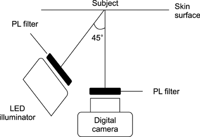
Fig. 1 A schematic diagram showing the arrangement of the equipment. LED: light emitting diode, PL: polarized light.
Reference
-
1. Kim DH, Choi JE, Ryu HJ, Seo SH, Kye YC, Ahn HH. Analytic parallel-polarized light imaging technique using various light-emitting diodes: a comparison with skin conductance values. Skin Res Technol. 2015; 21:158–163.
Article2. Kye H, Choi JE, Seo SH, Kye YC, Ahn HH. Application of analytic technique using green light parallel-polarized light images in various skin diseases. Ann Dermatol. 2016; 28:242–245.
Article3. Latreille J, Gardinier S, Ambroisine L, Mauger E, Tenenhaus M, Guéhenneux S, et al. Influence of skin colour on the detection of cutaneous erythema and tanning phenomena using reflectance spectrophotometry. Skin Res Technol. 2007; 13:236–241.
Article4. Rogers RS 3rd, Callen J, Wehr R, Krochmal L. Comparative efficacy of 12% ammonium lactate lotion and 5% lactic acid lotion in the treatment of moderate to severe xerosis. J Am Acad Dermatol. 1989; 21:714–716.
Article5. Wilkin J, Dahl M, Detmar M, Drake L, Liang MH, Odom R, et al. Standard grading system for rosacea: report of the national rosacea society expert committee on the classification and staging of rosacea. J Am Acad Dermatol. 2004; 50:907–912.
Article6. Taylor S, Westerhof W, Im S, Lim J. Noninvasive techniques for the evaluation of skin color. J Am Acad Dermatol. 2006; 54:5 Suppl 2. S282–S290.
Article7. Phillips SB, Kollias N, Gillies R, Muccini JA, Drake LA. Polarized light photography enhances visualization of inflammatory lesions of acne vulgaris. J Am Acad Dermatol. 1997; 37:948–952.
Article8. Farage MA. Enhancement of visual scoring of skin irritant reactions using cross-polarized light and parallel-polarized light. Contact Dermatitis. 2008; 58:147–155.
Article9. Ortonne JP, Gupta G, Ortonne N, Duteil L, Queille C, Mallefet P. Effectiveness of cross polarized light and fluorescence diagnosis for detection of sub-clinical and clinical actinic keratosis during imiquimod treatment. Exp Dermatol. 2010; 19:641–647.
Article10. Kollias N, Stamatas GN. Optical non-invasive approaches to diagnosis of skin diseases. J Investig Dermatol Symp Proc. 2002; 7:64–75.
Article11. Anderson RR. Polarized light examination and photography of the skin. Arch Dermatol. 1991; 127:1000–1005.
Article12. Muccini JA, Kollias N, Phillips SB, Anderson RR, Sober AJ, Stiller MJ, et al. Polarized light photography in the evaluation of photoaging. J Am Acad Dermatol. 1995; 33:765–769.
Article13. Bae EJ, Seo SH, Kye YC, Ahn HH. A quantitative assessment of the human skin surface using polarized light digital photography and its dermatologic significance. Skin Res Technol. 2010; 16:270–274.
Article14. Steinhoff M, Buddenkotte J, Aubert J, Sulk M, Novak P, Schwab VD, et al. Clinical, cellular, and molecular aspects in the pathophysiology of rosacea. J Investig Dermatol Symp Proc. 2011; 15:2–11.
Article
- Full Text Links
-
- Actions
-
Cited
- CITED
-
- Close
- Share
-
- Similar articles
-
- Immunohistochemical Profiling Reveals Distinct Inflammatory Landscape in Rosacea Subtypes
- A Study of the Clinical Features of Rosacea and a Comparison of Its Classifications
- A Rosacea Case Involving One Side of the Face Accompanied by Demodex Infestation: Unilateral Rosacea Fulminans
- Histological Findings in Korean Patients with Rosacea
- Clinical Evaluation of 168 Korean Patients with Rosacea: The Sun Exposure Correlates with the Erythematotelangiectatic Subtype
