Cancer Res Treat.
2017 Oct;49(4):890-897. 10.4143/crt.2016.325.
The Prognostic Values of Preoperative Tumor Volume and Tumor Diameter in T1N0 Papillary Thyroid Cancer
- Affiliations
-
- 1Division of Breast and Thyroid Surgical Oncology, Department of Surgery, St. Vincent's Hospital, College of Medicine, The Catholic University of Korea, Suwon, Korea. yjsuh@catholic.ac.kr
- KMID: 2394808
- DOI: http://doi.org/10.4143/crt.2016.325
Abstract
- PURPOSE
The current TNM staging system for papillary thyroid cancer (PTC), which is based on tumor diameter, may not precisely reflect the true tumor burden. Therefore, we investigated whether preoperative tumor volume might more accurately reflect tumor burden and predict prognosis in patients with T1N0 PTC than preoperative tumor diameter.
MATERIALS AND METHODS
We retrospectively reviewed data from 1,659 patients with T1N0 PTC, and after exclusion, a total of 1,081 patients were ultimately included. Tumor volume (V) was calculated for all patients using preoperative ultrasonography, and patients were grouped according to tumor diameter (T1a vs. T1b) and tumor volume (V1a vs. V1b). The recurrence-free survival (RFS) rates were then compared for these groups.
RESULTS
The mean follow-up time was 66.12±28.75 months, and 97.2% of the cohort experienced RFS. The optimal volume cut-off was defined as 0.545 cm³. There were no differences in RFS rates between T1a/T1b groups (all ages) and V1a/V1b groups (< 45 years of age). However, ≥ 45-year-old patients in the V1b group had a significantly poorer RFS rate than those in the V1a group. These results were confirmed by multivariate analysis.
CONCLUSION
Our results indicate that preoperative tumor volume may be more useful for predicting prognosis than tumor diameter in ≥ 45-year-old patients with T1N0 PTC.
Keyword
MeSH Terms
Figure
-
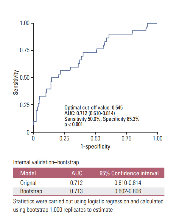
Fig. 1. Receiver operating characteristic (ROC) curve analysis to estimate the optimal cut-off tumor volume for predicting recurrence.
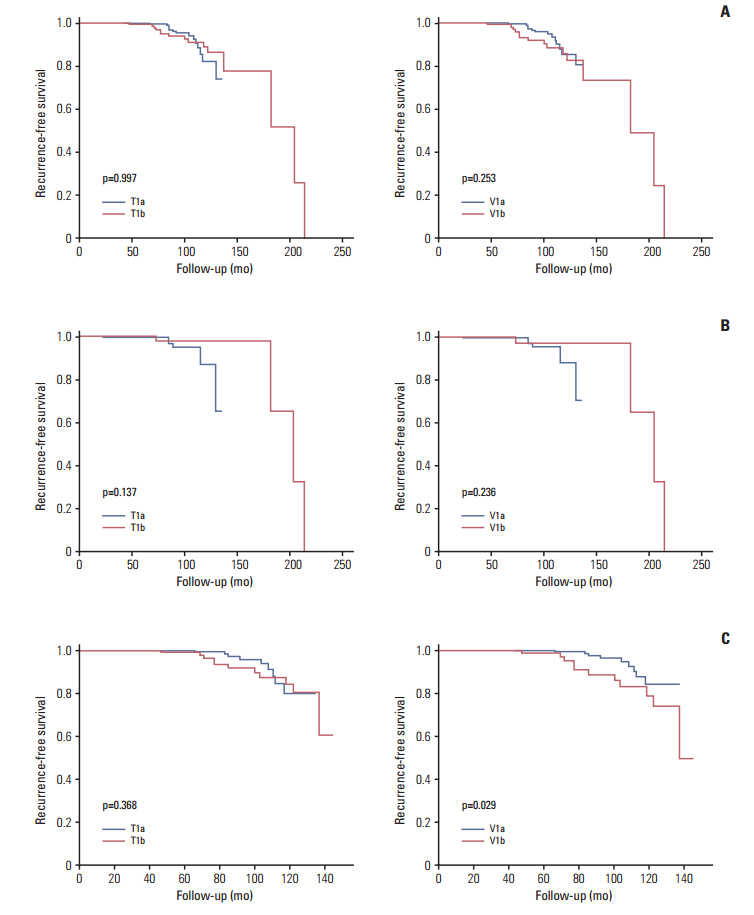
Fig. 2. Recurrence-free survival according to tumor diameter (T) and tumor volume categorization (V). Entire study population (A), patients aged < 45 years (B), and patients aged ≥ 45 years (C).
Cited by 1 articles
-
Prognostic influence of 3-dimensional tumor volume on breast cancer compared to conventional 1-dimensional tumor size
Ki-Tae Hwang, Wonshik Han, Sang Mok Lee, Jaewoo Choi, Jongjin Kim, Jiyoung Rhu, Young A Kim, Dong-Young Noh
Ann Surg Treat Res. 2018;95(4):183-191. doi: 10.4174/astr.2018.95.4.183.
Reference
-
References
1. Schlumberger MJ. Papillary and follicular thyroid carcinoma. N Engl J Med. 1998; 338:297–306.
Article2. DeSantis CE, Lin CC, Mariotto AB, Siegel RL, Stein KD, Kramer JL, et al. Cancer treatment and survivorship statistics, 2014. CA Cancer J Clin. 2014; 64:252–71.
Article3. Enewold L, Zhu K, Ron E, Marrogi AJ, Stojadinovic A, Peoples GE, et al. Rising thyroid cancer incidence in the United States by demographic and tumor characteristics, 1980-2005. Cancer Epidemiol Biomarkers Prev. 2009; 18:784–91.
Article4. Lee JH, Lee ES, Kim YS, Won NH, Chae YS. BRAF mutation and AKAP9 expression in sporadic papillary thyroid carcinomas. Pathology. 2006; 38:201–4.
Article5. Hall SF, Walker H, Siemens R, Schneeberg A. Increasing detection and increasing incidence in thyroid cancer. World J Surg. 2009; 33:2567–71.
Article6. Davies L, Welch HG. Increasing incidence of thyroid cancer in the United States, 1973-2002. JAMA. 2006; 295:2164–7.
Article7. Brito JP, Morris JC, Montori VM. Thyroid cancer: zealous imaging has increased detection and treatment of low risk tumours. BMJ. 2013; 347:f4706.
Article8. Ahn HS, Kim HJ, Welch HG. Korea's thyroid-cancer “epidemic”: screening and overdiagnosis. N Engl J Med. 2014; 371:1765–7.9. Edge SB, Byrd DR, Compton CC, Fritz AG, Greene FL, Trotti A. AJCC cancer staging manual. 7th ed. New York: Springer;2010.10. Haugen BR, Alexander EK, Bible KC, Doherty GM, Mandel SJ, Nikiforov YE, et al. 2015 American Thyroid Association management guidelines for adult patients with thyroid nodules and differentiated thyroid cancer: The American Thyroid Association Guidelines Task Force on Thyroid Nodules and Differentiated Thyroid Cancer. Thyroid. 2016; 26:1–133.
Article11. Tsai CH, Lin CM, Hsieh CC, Hsu WH, Wang HW, Wang LS. Tumor volume is a better prognostic factor than greatest tumor diameter in stage Ia non-small cell lung cancer. Thorac Cardiovasc Surg. 2006; 54:537–43.
Article12. Davis KS, Lim CM, Clump DA, Heron DE, Ohr JP, Kim S, et al. Tumor volume as a predictor of survival in human papillomavirus-positive oropharyngeal cancer. Head Neck. 2016; 38 Suppl 1:E1613–7.
Article13. Park KN, Kang KY, Hong HS, Jeong HS, Lee SW. Predictive value of estimated tumor volume measured by ultrasonography for occult central lymph node metastasis in papillary thyroid carcinoma. Ultrasound Med Biol. 2015; 41:2849–54.
Article14. Anderson KL Jr, Youngwirth LM, Scheri RP, Stang MT, Roman SA, Sosa JA. T1a versus T1b differentiated thyroid cancers: do we need to make the distinction? Thyroid. 2016; 26:1046–52.
Article15. Wang LY, Nixon IJ, Palmer FL, Thomas D, Tuttle RM, Shaha AR, et al. Comparable outcomes for patients with pT1a and pT1b differentiated thyroid cancer: Is there a need for change in the AJCC classification system? Surgery. 2014; 156:1484–9.
Article16. Chereau N, Tresallet C, Noullet S, Godiris-Petit G, Tissier F, Leenhardt L, et al. Does the T1 subdivision correlate with the risk of recurrence of papillary thyroid cancer? Langenbecks Arch Surg. 2016; 401:223–30.
Article17. Moon HJ, Yoon JH, Kwak JY, Chung WY, Nam KH, Jeong JJ, et al. Positive predictive value and interobserver variability of preoperative staging sonography for thyroid carcinoma. AJR Am J Roentgenol. 2011; 197:W324–30.
Article18. Park JS, Son KR, Na DG, Kim E, Kim S. Performance of preoperative sonographic staging of papillary thyroid carcinoma based on the sixth edition of the AJCC/UICC TNM classification system. AJR Am J Roentgenol. 2009; 192:66–72.
Article
- Full Text Links
-
- Actions
-
Cited
- CITED
-
- Close
- Share
-
- Similar articles
-
- Clinical implications of preoperative thyrotropin serum concentrations in patients who underwent thyroidectomy for nonfunctioning nodule(s)
- Expression of Vascular Endothelial Growth Factor (VEGF) in Papillary Thyroid Carcinoma
- Prognostic Factors for Locally Invasive Papillary Thyroid Carcinomas
- Personalized Follow Up in Thyroid Cancer Using Molecular Markers
- Medullary and Papillary Thyroid Carcinoma as a Collision Tumor: Report of Five Cases
