J Korean Soc Radiol.
2017 Nov;77(5):333-338. 10.3348/jksr.2017.77.5.333.
CT Findings of Immunoglobulin G4 Related Periaortitis and Periarterities: A Case Report
- Affiliations
-
- 1Department of Radiology, Chungnam National University Hospital, Chungnam National University School of Medicine, Daejeon, Korea. haneul88@hanmail.net
- 2Department of Pathology, Chungnam National University Hospital, Chungnam National University School of Medicine, Daejeon, Korea.
- 3Division of Cardiology, Department of Internal Medicine, Chungnam National University Hospital, Chungnam National University School of Medicine, Daejeon, Korea.
- KMID: 2394048
- DOI: http://doi.org/10.3348/jksr.2017.77.5.333
Abstract
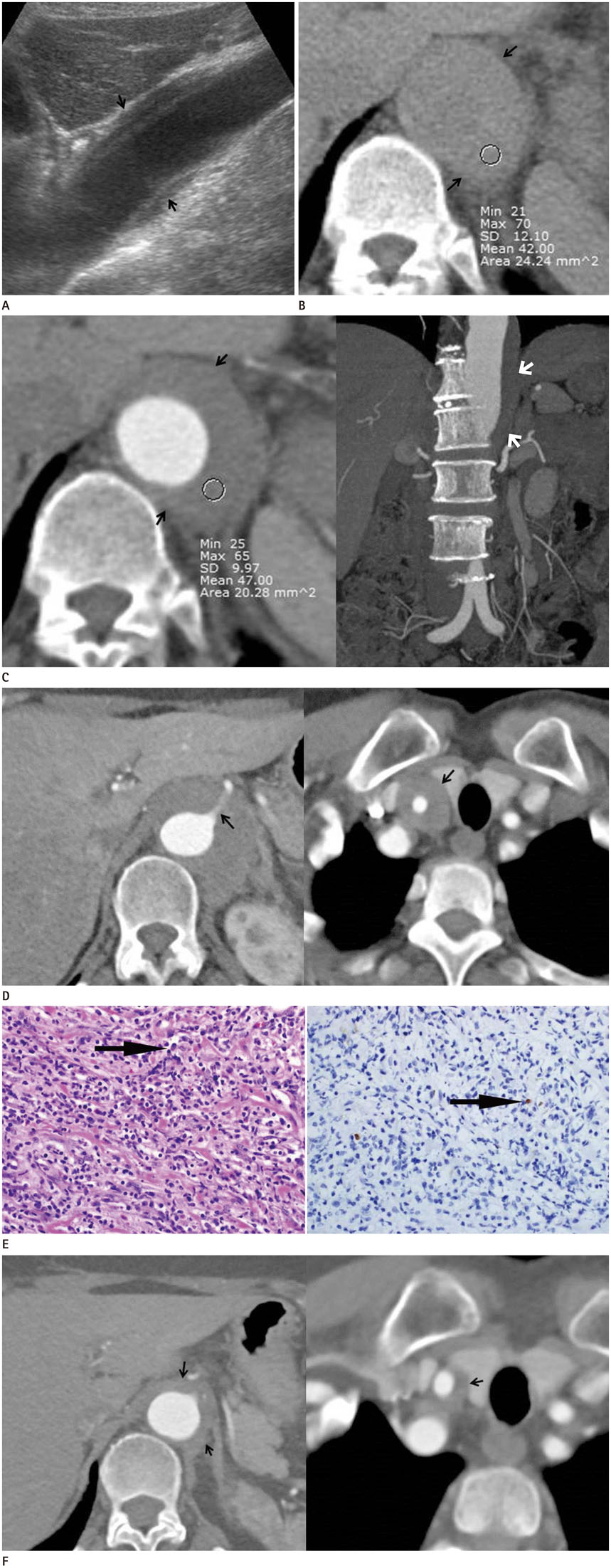
- Immunoglobulin G4 (IgG4)-related periaortitis and periarteritis are rare systemic inflammatory and fibrosclerosing diseases, usually involving the aorta and its main branches. We report a pathologically confirmed case of IgG4-related periaortitis involving the thoracoabdominal aorta, which can be confused with intramural hematoma or periaortic lymphoma.
MeSH Terms
Figure
Reference
-
1. Lipton S, Warren G, Pollock J, Schwab P. IgG4-related disease manifesting as pachymeningitis and aortitis. J Rheumatol. 2013; 40:1236–1238.2. Stone JH. L45. Aortitis, retroperitoneal fibrosis, and IgG4-related disease. Presse Med. 2013; 42(4 Pt 2):622–625.3. Mizushima I, Inoue D, Yamamoto M, Yamada K, Saeki T, Ubara Y, et al. Clinical course after corticosteroid therapy in IgG4-related aortitis/periaortitis and periarteritis: a retrospective multicenter study. Arthritis Res Ther. 2014; 16:R156.4. Babur Güler G, Cantürk E, Güler E, Oran G, Demir GG, Akçevin A, et al. IgG4-related aortitis mimicking intramural hematoma. Anatol J Cardiol. 2016; 16:728–729.5. Inoue D, Zen Y, Abo H, Gabata T, Demachi H, Yoshikawa J, et al. Immunoglobulin G4-related periaortitis and periarteritis: CT findings in 17 patients. Radiology. 2011; 261:625–633.6. Siddiquee Z, Smith RN, Stone JR. An elevated IgG4 response in chronic infectious aortitis is associated with aortic atherosclerosis. Mod Pathol. 2015; 28:1428–1434.7. Nishimura S, Amano M, Izumi C, Kuroda M, Yoshikawa Y, Takahashi Y, et al. Multiple coronary artery aneurysms and thoracic aortitis associated with IgG4-related disease. Intern Med. 2016; 55:1605–1609.8. Zambetti BR, Garrett E Jr. Plasmacytic aortitis with occlusion of the right coronary artery. Am J Case Rep. 2016; 17:549–552.9. Tran MN, Langguth D, Hart G, Heiner M, Rafter A, Fleming SJ, et al. IgG4-related systemic disease with coronary arteritis and aortitis, causing recurring critical coronary ischemia. Int J Cardiol. 2015; 201:33–34.10. Rousselin C, Pontana F, Puech P, Lambert M. [Differential diagnosis of aortitis]. Rev Med Interne. 2016; 37:256–263.
- Full Text Links
- Actions
-
Cited
- CITED
-
- Close
- Share
- Similar articles
-
- A Case of IgG4-Related Disease with Pachymeningitis and Periaortitis
- Non-IgG4-Related Fibrosclerosing Periaortitis with Multisystemic Involvement
- Unusual Manifestation of Immunoglobulin G4-Related Disease Involving the Retroperitoneum: A Case Report
- A Rare Case of Granulomatosis with Polyangiitis-Related Periaortitis at the Ascending Aorta
- Immunoglobulin G4-Related Lung Disease Mimicking Lung Cancer: Two Case Reports


