Feasibility of Fabricating Variable Density Phantoms Using 3D Printing for Quality Assurance (QA) in Radiotherapy
- Affiliations
-
- 1Department of Radiation Oncology, Yeungnam University Medical Center, Daegu, Korea. skkim3@ynu.ac.kr
- 2Gyeongnam Science High School, Jinju, Korea.
- KMID: 2393371
- DOI: http://doi.org/10.14316/pmp.2017.28.3.106
Abstract
- The variable density phantom fabricated with varying the infill values of 3D printer to provide more accurate dose verification of radiation treatments. A total of 20 samples of rectangular shape were fabricated by using the Finebot™ (AnyWorks; Korea) Z420 model (width×length×height=50 mm×50 mm×10 mm) varying the infill value from 5% to 100%. The samples were scanned with 1-mm thickness using a Philips Big Bore Brilliance CT Scanner (Philips Medical, Eindhoven, Netherlands). The average Hounsfield Unit (HU) measured by the region of interest (ROI) on the transversal CT images. The average HU and the infill values of the 3D printer measured through the 2D area profile measurement method exhibited a strong linear relationship (adjusted R-square=0.99563) in which the average HU changed from −926.8 to 36.7, while the infill values varied from 5% to 100%. This study showed the feasibility fabricating variable density phantoms using the 3D printer with FDM (Fused Deposition Modeling)-type and PLA (Poly Lactic Acid) materials.
Figure
-
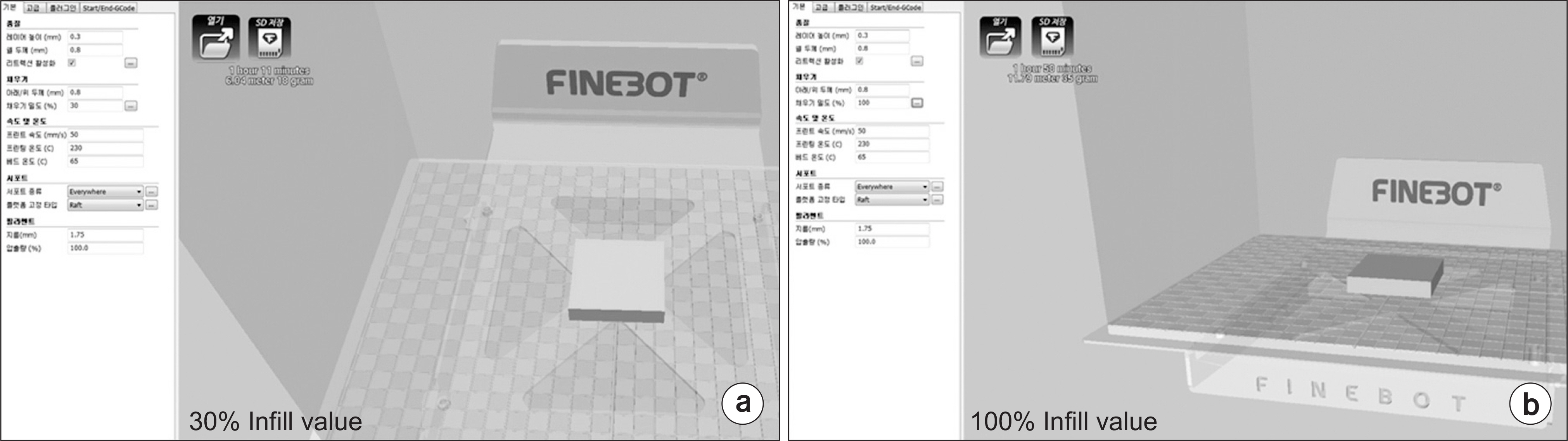
Fig. 1. A total of 20 samples with dimensions (width×length×height=50 mm×50 mm×10 mm) were fabricated for infill values ranging from 5% to 100% by using FinebotTM (AnyWorks; Korea) Z420 3D printer model.
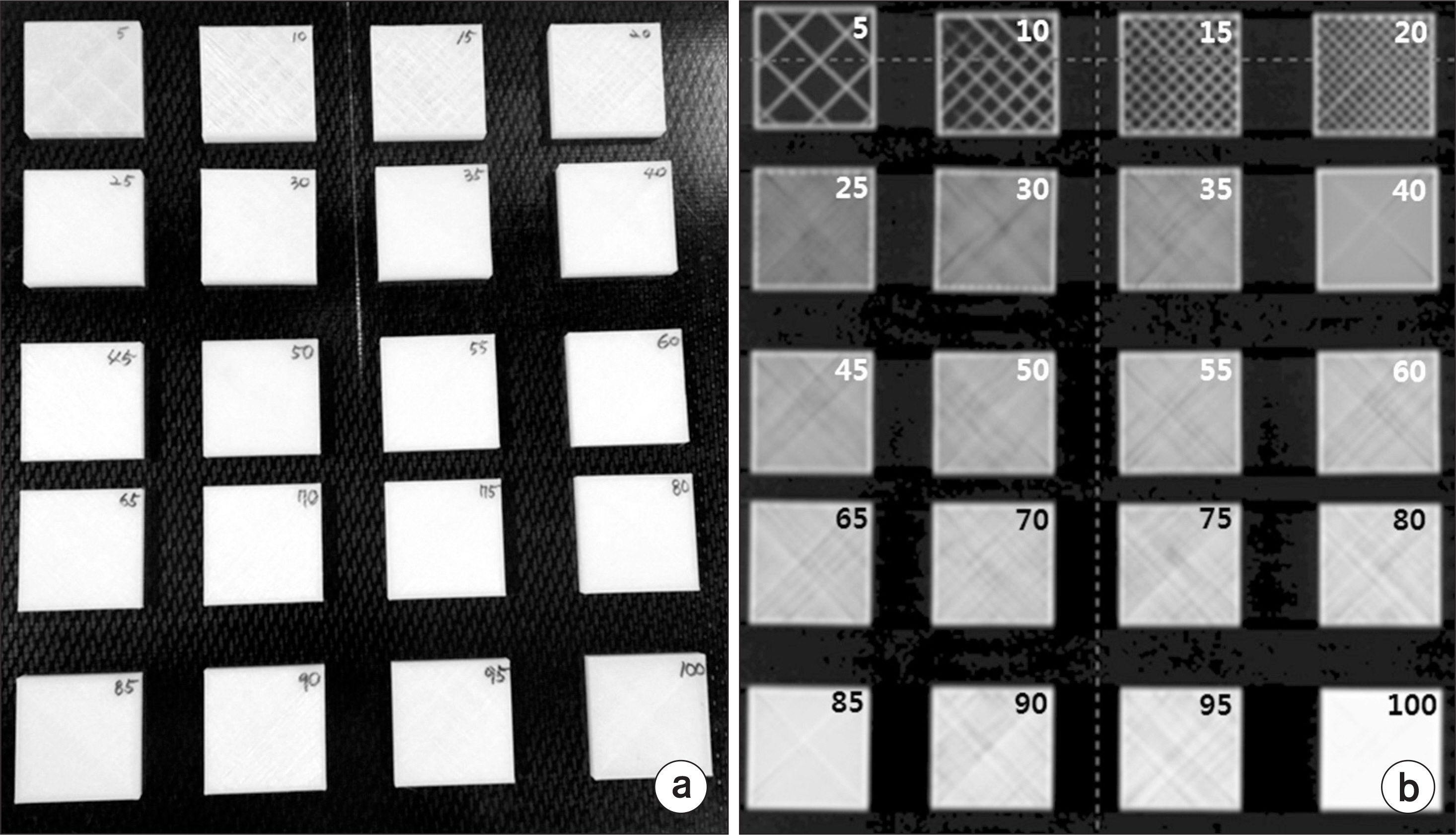
Fig. 2. (a) Photograph of samples with infill values from 5% to 100%, (b) Frontal image of samples with infill values from 5% to 100% obtained by CT scan.

Fig. 3. (a) HU was measured by using a two-dimensional area profile function at the center of the sample on a transversal image by CT scan. (b) Horizontal ROI Profile, vertical ROI Profile, pixel statistics, and ROI for the measured area of the sample with infill value of 5%.
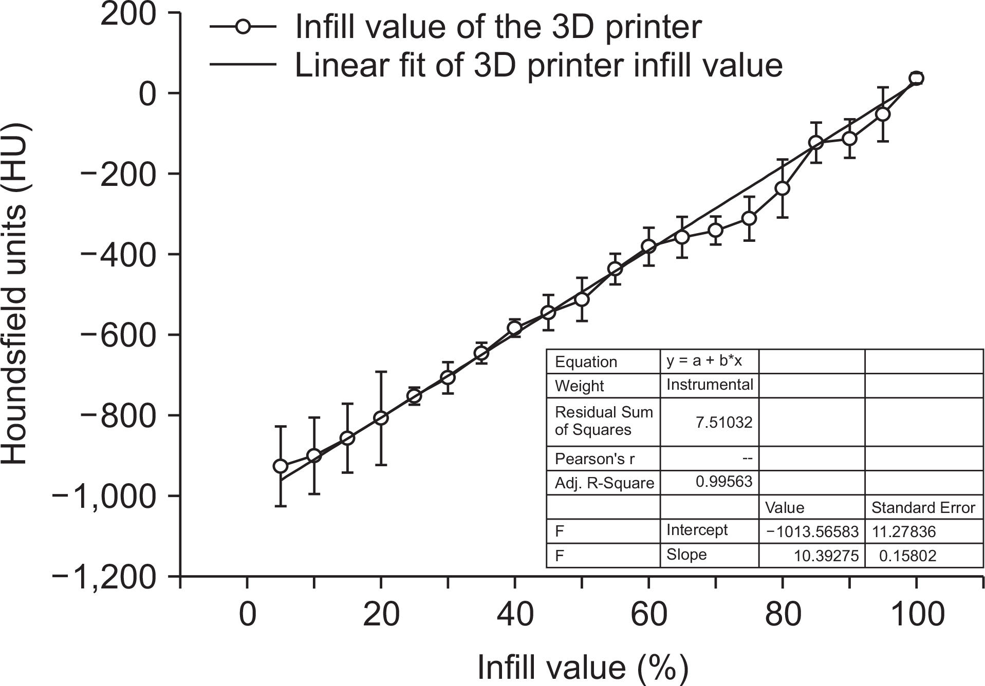
Fig. 4. Correlation between infill value (%) and Hounsfield unit (HU) of the 3D printer.
Cited by 1 articles
-
Radiological Characteristics of Materials Used in 3-Dimensional Printing with Various Infill Densities
So-Yeon Park, Noorie Choi, Byeong Geol Choi, Dong Myung Lee, Na Young Jang
Prog Med Phys. 2019;30(4):155-159. doi: 10.14316/pmp.2019.30.4.155.
Reference
-
1. Oh SA, Kang MK, Yea JW, Kim SK, Oh YK. Study of the penumbra for high-energy photon beams with Gafchromic TM EBT2 films. Journal of the Korean Physical Society. 2012; 60:1973–76.2. Khan FM, Gibbons JP. Khan's the physics of radiation therapy: Lippincott Williams & Wilkins. 2014.3. Ezzell GA, Burmeister JW, Dogan N, LoSasso TJ, Mecha-lakos JG, et al. IMRT commissioning: multiple institution planning and dosimetry comparisons, a report from AAPM Task Group 119. Medical physics. 2009; 36:5359–73.
Article4. Yea JW, Park JW, Kim SK, Kim DY, Kim JG, et al. Feasibility of a 3D-printed anthropomorphic patient-specific head phantom for patient-specific quality assurance of intensity-modulated radiotherapy. PloS one. 2017; 12:e0181560.
Article5. Oh SA, Kim SK, Kang MK, Yea JW, Kim EC. Dosimetric verification of enhanced dynamic wedges by a 2D ion chamber array. Journal of the Korean Physical Society. 2013; 63:2215–19.
Article6. Oh SA, Yea JW, Lee R, Park HB, Kim SK. Dosimetric Verifications of the Output Factors in the Small Field Less Than 3 cm2 Using the Gafchromic EBT2 Films and the Various Detectors. Progress in Medical Physics. 2014; 25:218–24.7. Kim SK, Kang MK, Yea JW, Oh SA. Dosimetric evaluation of a moving tumor target in intensity-modulated radiation therapy (IMRT) for lung cancer patients. Journal of the Korean Physical Society. 2013; 63:67–70.
Article8. Oh SA, Kang MK, Yea JW, Kim SH, Kim KH, et al. Comparison of intensity modulated radiation therapy dose calculations with a PBC and AAA algorithms in the lung cancer. Korean J Med Phys. 2012; 23:48–53.9. Bush K, Gagne I, Zavgorodni S, Ansbacher W, Beckham W. Dosimetric validation of Acuros® XB with Monte Carlo methods for photon dose calculations. Medical physics. 2011; 38:2208–21.10. Fogliata A, Nicolini G, Clivio A, Vanetti E, Cozzi L. Critical appraisal of Acuros XB and Anisotropic Analytic Algorithm dose calculation in advanced non-small-cell lung cancer treatments. International Journal of Radiation Oncology∗ Biology∗ Physics. 2012; 83:1587–95.
Article11. Tsuruta Y, Nakata M, Nakamura M, Matsuo Y, Higashi-mura K, et al. Dosimetric comparison of Acuros XB, AAA, and XVMC in stereotactic body radiotherapy for lung cancer. Medical physics. 2014; 41.
Article12. Ehler ED, Barney BM, Higgins PD, Dusenbery KE. Patient specific 3D printed phantom for IMRT quality assurance. Physics in medicine and biology. 2014; 59:5763.
Article13. Madamesila J, McGeachy P, Barajas JEV, Khan R. Characterizing 3D printing in the fabrication of variable density phantoms for quality assurance of radiotherapy. Physica Medica. 2016; 32:242–7.
Article14. Park JW, Oh SA, Yea JW, Kang MK. Fabrication of malleable three-dimensional-printed customized bolus using three-dimensional scanner. PloS one. 2017; 12:e0177562.
Article15. Park J, Yea J. Three-dimensional customized bolus for intensity-modulated radiotherapy in a patient with Kimura's disease involving the auricle. Cancer/Radio-thérapie. 2016; 20:205–9.
Article
- Full Text Links
-
- Actions
-
Cited
- CITED
-
- Close
- Share
-
- Similar articles
-
- Use of Statistical Process Control for Quality Assurance in Radiation Therapy
- Clinical Implementation of 3D Printing in the Construction of Patient Specific Bolus for Photon Beam Radiotherapy for Mycosis Fungoides
- Analysis on the Dosimetric Characteristics of Tangential Breast Intensity Modulated Radiotherapy
- Clinical Applications of Three-Dimensional Printing in Cardiovascular Disease
- The Development of Quality Assurance Program for CyberKnife
