Comparison of Short-term Clinical Outcomes between Scleral Fixation vs. Iris Fixation of Dislocated IOL
- Affiliations
-
- 1Department of Ophthalmology, Yeouido St. Mary's Hospital, College of Medicine, The Catholic University of Korea, Seoul, Korea. sara514@catholic.ac.kr
- KMID: 2393164
- DOI: http://doi.org/10.3341/jkos.2017.58.10.1131
Abstract
- PURPOSE
To compare clinical outcomes between iris fixation and scleral fixation as treatments for dislocated Intra Ocular Lens.
METHODS
Ten eyes of 10 patients underwent scleral fixation (scleral fixation group) and 8 eyes of 8 patients underwent iris fixation (iris fixation group) were enrolled in this retrospective study. In each group, visual acuity and intra ocular pressure, slit lamp examination, fundus examination, refraction, keratometry, axial length and anterior chamber depth were measured before the surgery. Regular follow up was made 1 day, 1 week, 1 month, and 2 months after surgery and visual acuity, intra ocular pressure, slit lamp exam, refractory error, anterior chamber depth, intraocular lens (IOL) tilting, and decentration were measured at each visit.
RESULTS
There were no significant differences in uncorrected visual acuity (UCVA), best corrected visual acuity (BCVA), and refractive error for patients with iris and scleral fixation before and after surgery. Patients with iris fixation had significantly deeper anterior chamber depth (ACD) and more IOL tilting than patients with scleral fixation.
CONCLUSIONS
In this study, the iris fixation group tended to have more IOL tilting and deepening of anterior chamber depth than the scleral fixation group. We can use this information to choose the appropriate surgical method for dislocated IOL and to select of new IOL.
Keyword
MeSH Terms
Figure
-
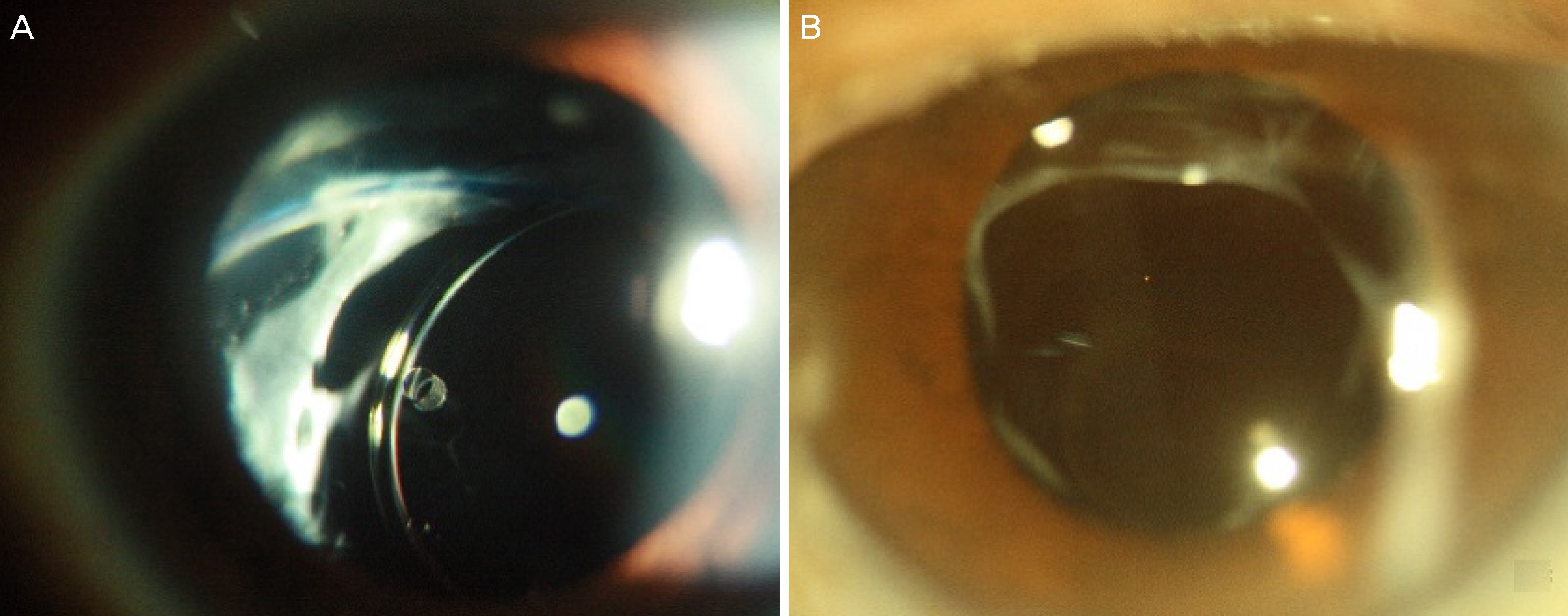
Figure 1. Representative images of dislocated intraocular lens. (A) Inferonasally tilted lens (B) Inferiorly tilted lens.
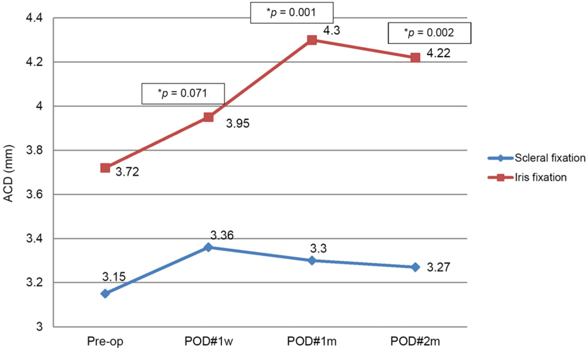
Figure 2. Changes of Anterior chamber depth (ACD) over time after Scleral or Iris fixation of intraocular lens (mm). Patients with iris fixation had significantly deeper ACD than patients with scleral fixation. Pre-op. = pre-operative; POD = post operative day; w = week; m = month(s). * Values which are statistically significant.

Figure 3. Changes of intraocular lens (IOL) tilting (°) over time after Scleral or Iris fixation of IOL. Patients with iris fixation had more IOL tilting than patients with scleral fixation. POD = post operative day; w = week; m = month(s). * Values which are statistically significant.
Cited by 1 articles
-
Short-term Clinical Outcomes of Scleral Fixation of Intraocular Lenses Using a Scleral Tunnel and Groove
Tae Kyu Moon, Jae Yong Jang, Hyun Ho Jung, Yong Sok Ji
J Korean Ophthalmol Soc. 2019;60(3):246-252. doi: 10.3341/jkos.2019.60.3.246.
Reference
-
References
1. Gross JG, Kokame GT, Weinberg DV; Dislocated In-The-Bag Intraocular Lens Study Group. In-the-bag intraocular lens dislocation. Am J Ophthalmol. 2004; 137:630–5.2. Smiddy WE, Ibanez GV, Alfonso E, Flynn HW Jr. Surgical abdominal of dislocated intraocular lenses. J Cataract Refract Surg. 1995; 21:64–9.3. Wagoner MD, Cox TA, Ariyasu RG, et al. Intraocular lens abdominalation in the absence of capsular support: a report by the American Academy of Ophthalmology. Ophthalmology. 2003; 110:840–59.4. Krė pštė L, Kuzmienė L, Miliauskas A, Janulevič ienė I. Possible predisposing factors for late intraocular lens dislocation after abdominal cataract surgery. Medicina (Kaunas). 2013; 49:229–34.5. Fernández-Buenaga R, Alio JL, Pérez-Ardoy AL, et al. Late in-the-bag intraocular lens dislocation requiring explantation: risk factors and outcomes. Eye (Lond). 2013; 27:795–801. quiz 802.
Article6. Davis D, Brubaker J, Espandar L, et al. Late in-the-bag abdominal intraocular lens dislocation: evaluation of 86 consecutive cases. Ophthalmology. 2009; 116:664–70.7. Masket S, Osher RH. Late complications with intraocular lens abdominal after capsulorhexis in pseudoexfoliation syndrome. J Cataract Refract Surg. 2002; 28:1481–4.8. Brod RD, Flynn Jr HW, Clarkson JG, Blankenship GW. Management options for retinal detachment in the presence of a posteriorly dislocated intraocular lens. Retina. 1990; 10:50–6.
Article9. Gimbel HV, Condon GP, Kohnen T, et al. Late in-the-bag abdominal lens dislocation: incidence, prevention, and management. J Cataract Refract Surg. 2005; 31:2193–204.10. Lim MC, Doe EA, Vroman DT, et al. Late onset lens particle abdominal as a consequence of spontaneous dislocation of an abdominal lens in pseudoexfoliation syndrome. Am J Ophthalmol. 2001; 132:261–3.11. Kim KH, Kim WS. Comparison of clinical outcomes of iris fixation and scleral fixation as treatment for intraocular lens dislocation. Am J Ophthalmol. 2015; 160:463–9. e1.
Article12. Scharioth GB, Prasad S, Georgalas I, et al. Intermediate results of sutureless intrascleral posterior chamber intraocular lens fixation. J Cataract Refract Surg. 2010; 36:254–9.
Article13. Garcia-Rojas L, Paulin-Huerta JM, Chavez-Mondragon E, Ramirez-Miranda A. Intraocular lens iris fixation. Clinical and macular OCT outcomes. BMC Res Notes. 2012; 5:560.
Article14. Lyle W, Jin JC. Secondary intraocular lens implantation: anterior chamber vs posterior chamber lenses. Ophthalmic Surg. 1993; 24:375–81.
Article15. Hall JR, Muenzler WS. Intraocular lens replacement in abdominal bullous keratopathy. Trans Ophthalmol Soc U K. 1985; 104(Pt 5):541–5.16. Michaeli A, Soiberman U, Loewenstein A. Outcome of iris abdominal of subluxated intraocular lenses. Graefes Arch Clin Exp Ophthalmol. 2012; 250:1327–32.17. Engren AL, Behndig A. Anterior chamber depth, intraocular lens position, and refractive outcomes after cataract surgery. J Cataract Refract Surg. 2013; 39:572–7.
Article18. Sasaki K, Sakamoto Y, Shibata T, et al. Measurement of abdominal intraocular lens tilting and decentration using Scheimpflug images. J Cataract Refract Surg. 1989; 15:454–7.19. Hayashi K, Hayashi H, Nakao F, Hayashi F. Intraocular lens tilt and decentration, anterior chamber depth, and refractive error after abdominal suture fixation surgery. Ophthalmology. 1999; 106:878–82.20. Drexler W, Findl O, Menapace R, et al. Partial coherence abdominal: a novel approach to biometry in cataract surgery. Am J Ophthalmol. 1998; 126:524–34.21. Hayashi K, Harada M, Hayashi H, et al. Decentration and tilt of polymethyl methacrylate, silicone, and acrylic soft intraocular lenses. Ophthalmology. 1997; 104:793–8.
Article
- Full Text Links
-
- Actions
-
Cited
- CITED
-
- Close
- Share
-
- Similar articles
-
- Comparison of Short-term Clinical Outcomes between Sutured Scleral Fixation and Modified Yamane Sutureless Scleral Fixation
- Four-flanged Technique for Scleral Fixation of a Dislocated Four-eyelet Intraocular Lens
- Scleral Fixation of Intraocular Lens and Retropupillary Fixation of Iris Claw Lens for Aphakic Eyes
- Outcomes of Re-fixation after the First Intraocular Lens Scleral Fixation
- Astigmatic Changes and Clinical Outcomes after Scleral Fixation of IOL
