Cerebellar Liponeurocytoma: Relevant Clinical Cytogenetic Findings
- Affiliations
-
- 1Department of Neurosurgery, University of California-Los Angeles, Los Angeles, CA, USA. atucker@mednet.ucla.edu
- 2Division of Neuropathology, Department of Pathology and Laboratory Medicine, University of California-Los Angeles, Los Angeles, CA, USA.
- 3Division of Neuroradiology, Department of Radiological Sciences, University of California-Los Angeles, Los Angeles, CA, USA.
- 4Division of Clinical and Molecular Cytogenetics, Department of Pathology and Laboratory Medicine, University of California-Los Angeles, Los Angeles, CA, USA.
- KMID: 2392603
- DOI: http://doi.org/10.4132/jptm.2016.07.24
Abstract
- No abstract available.
MeSH Terms
Figure
-
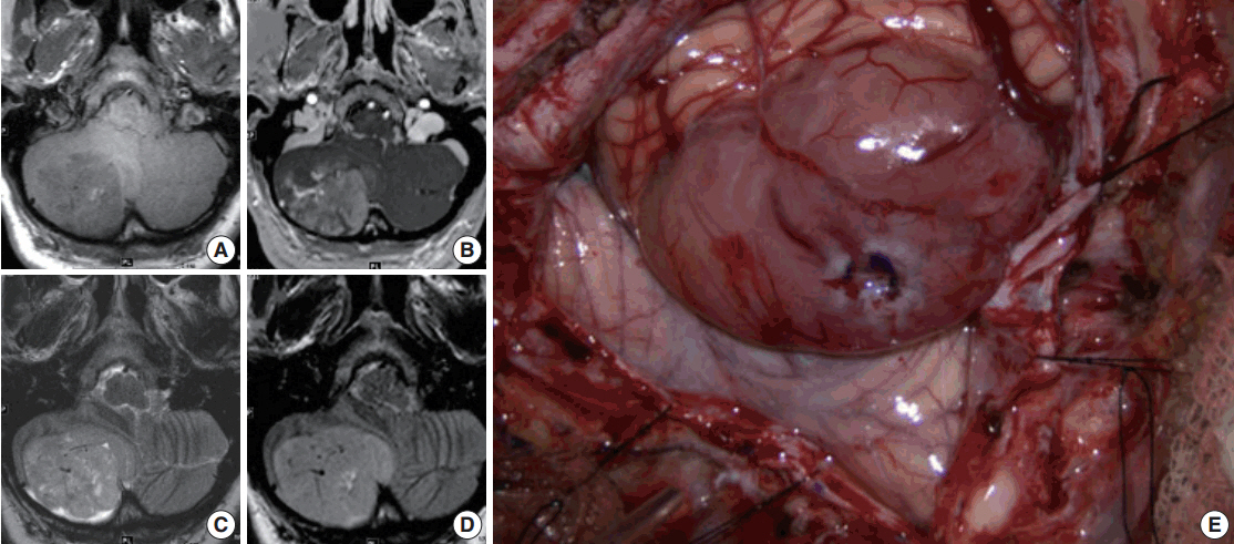
Fig. 1. Radiographic and gross appearance. Magnetic resonance imaging of heterogeneously-enhancing right cerebellar hemisphere mass. Axial T1-weighted precontrast (A), T1-weighted postcontrast (B), T2-weighted (C), and fluid attenuated inversion recovery (D) images through the posterior fossa. Intraoperative photograph of tumor and surrounding cerebellum (E).
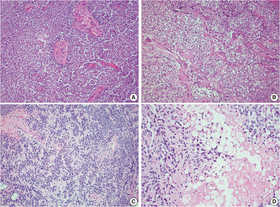
Fig. 2. Hematoxylin and eosin staining. (A) Areas of dense cellularity and scattered prominent vascular channels. (B) Oligodendroglial-like areas demonstrating cells with distinct perinuclear halos and a chicken wire vascular pattern. (C) Region of palisading tumor nuclei reminiscent of ependymoma. (D) Foci of lipidized cells.
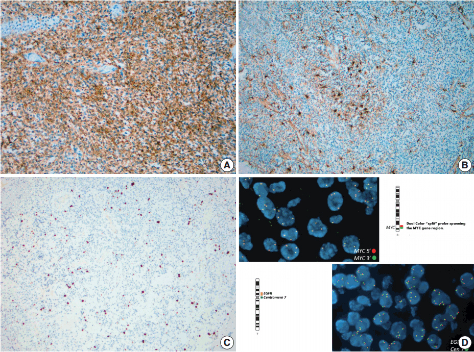
Fig. 3. Immunohistochemistry and fluorescent in-situ hybridization. (A) Diffuse synaptophysin immunoreactivity within the tumor. (B) Reactive gliosis on glial fibrillary acidic protein immunohistochemistry, but no tumor immunoreactivity. (C) Low Ki-67 nuclear labeling index, estimated at 3%–5%. (D) Fluorescent in-situ hybridization demonstrating lack of epidermal growth factor receptor (EGFR) amplification or MYC rearrangement.
Reference
-
1. Bechtel JT, Patton JM, Takei Y. Mixed mesenchymal and neuroectodermal tumor of the cerebellum. Acta Neuropathol. 1978; 41:261–3.
Article2. Nishimoto T, Kaya B. Cerebellar liponeurocytoma. Arch Pathol Lab Med. 2012; 136:965–9.
Article3. George DH, Scheithauer BW. Central liponeurocytoma. Am J Surg Pathol. 2001; 25:1551–5.
Article4. Louis DN, Ohgaki H, Wiestler OD, et al. The 2007 WHO classification of tumours of the central nervous system. Acta Neuropathol. 2007; 114:97–109.
Article5. Jenkinson MD, Bosma JJ, Du Plessis D, et al. Cerebellar liponeurocytoma with an unusually aggressive clinical course: case report. Neurosurgery. 2003; 53:1425–7.
Article6. Gonzalez-Campora R, Weller RO. Lipidized mature neuroectodermal tumour of the cerebellum with myoid differentiation. Neuropathol Appl Neurobiol. 1998; 24:397–402.7. Oudrhiri MY, Raouzi N, El Kacemi I, et al. Understanding cerebellar liponeurocytomas: case report and literature review. Case Rep Neurol Med. 2014; 2014:186826.
Article8. Taruscio D, Danesi R, Montaldi A, Cerasoli S, Cenacchi G, Giangaspero F. Nonrandom gain of chromosome 7 in central neurocytoma: a chromosomal analysis and fluorescence in situ hybridization study. Virchows Arch. 1997; 430:47–51.
Article9. Rodriguez FJ, Mota RA, Scheithauer BW, et al. Interphase cytogenetics for 1p19q and t(1;19)(q10;p10) may distinguish prognostically relevant subgroups in extraventricular neurocytoma. Brain Pathol. 2009; 19:623–9.
Article10. Horstmann S, Perry A, Reifenberger G, et al. Genetic and expression profiles of cerebellar liponeurocytomas. Brain Pathol. 2004; 14:281–9.
Article
- Full Text Links
-
- Actions
-
Cited
- CITED
-
- Close
- Share
-
- Similar articles
-
- Cerebellar Liponeurocytoma with an Unusually Aggressive Histopathology : Case Report and Review of the Literature
- A Cerebellar Infarction Presented with a Clinical Seizure
- Chromosome Analysis of Ascitic Fluids from Patients with Malignant Tumor
- A Case of Cavernous Angioma of the Cerebellar Vermis
- Neuro-otological aspects of cerebellar stroke
