Perinatology.
2017 Sep;28(3):92-95. 10.14734/PN.2017.28.3.92.
Successful Isolation and Treatment of Cogenital Tuberculosis Infection Occurred in Premature Infants in the Neonatal Intensive Care Unit
- Affiliations
-
- 1Department of Pediatric, Hallym University Kangnam Sacred Heart Hospital, Seoul, Korea. neosung@hallym.or.kr
- 2Division of Infectious Diseases, Department of Internal Medicine, Hallym University Kangnam Sacred Heart Hospital, Seoul, Korea.
- KMID: 2392429
- DOI: http://doi.org/10.14734/PN.2017.28.3.92
Abstract
- Congenital tuberculosis is a very rare disease and the affected infants are often delivered prematurely. Even though proper treatment, the mortality rate is high. The preterm male infant born at 24 weeks of gestational age with birth weight of 760 g was admitted to neonatal intensive care unit (NICU) for intensive treatment. One month after delivery, his mother visited to outpatient clinic for headache and nausea and later she was diagnosed with tuberculous meningitis. Then, the preterm infant who was already hospitalized was examined for the possibility of congenital infection. As a result, congenital tuberculosis infection was confirmed by positive reaction with tuberculosis in acid-fast bacilli stain, culture and polymerase chain reaction. The infant was completely isolated for 2 weeks and anti-tuberculosis drugs were administrated. We performed chest radiography and skin reaction test for the other 29 infants and 60 co-workers in the NICU during the same period. The infants were treated with prophylactic antituberculous drug. Three months later, no abnormal findings were observed in any infants and any co-workers during the follow up period. We report the experience of successful treatment and isolation for congenital tuberculosis in premature infants.
MeSH Terms
Figure
-
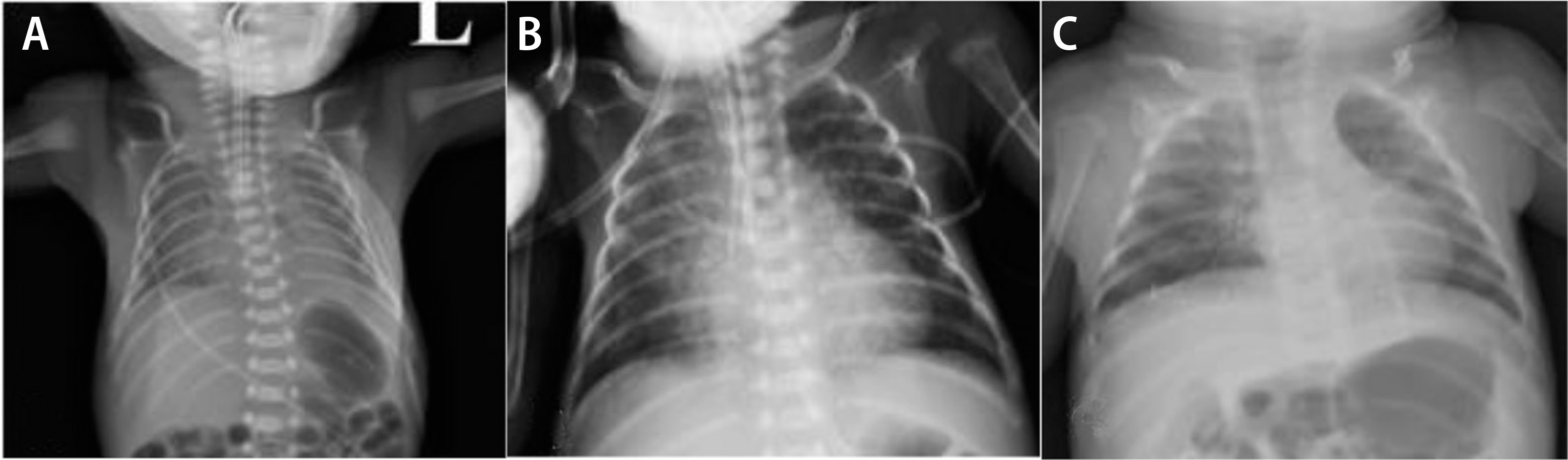
Fig. 1 Chest x-ray of infant. (A) Chest X-ray at hospital day 1 showed suspicious of respiratory distress syndrome. (B) Chest X-ray at hospital day 40 taken when the infant's mother diagnosed with tuberculosis meningitis. (C) Chest x-ray at hospital day 140 showed bronchopulmonary dysplasia.
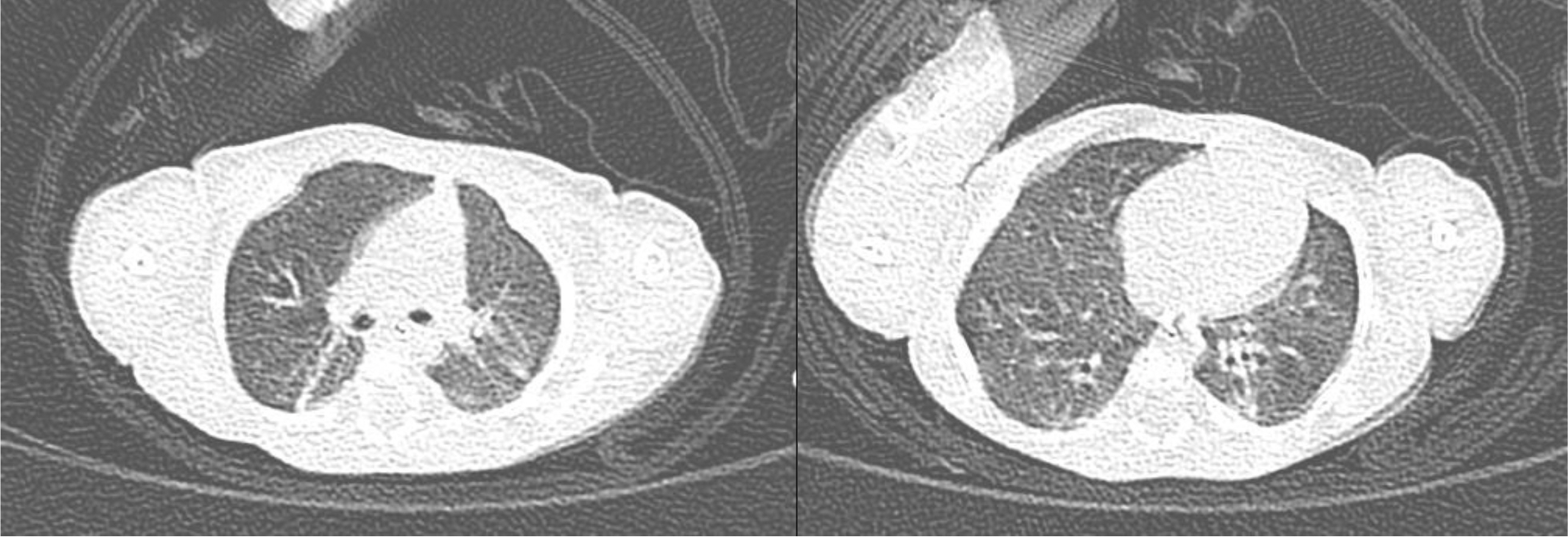
Fig. 2 Chest computed tomography of the infants taken before discharge, i.e., hospital days of 138. Several linear atelectasis in both lungs was shown in mostly dependent portion.
Reference
-
1). Kitai I., Morris SK., Kordy F., Lam R. Diagnosis and management of pediatric tuberculosis in Canada. CMAJ. 2017. 189:E11–6.
Article2). Schaaf HS., Collins A., Bekker A., Davies PD. Tuberculosis at extremes of age. Respirology. 2010. 15:747–63.
Article3). Ahn JG., Kim DS., Kim KH. Nosocomial exposure to active pulmonary tuberculosis in a neonatal intensive care unit. Am J Infect Control. 2015. 43:1292–5.
Article4). Skevaki CL., Kafetzis DA. Tuberculosis in neonates and infants: epidemiology, pathogenesis, clinical manifestations, diagnosis, and management issues. Paediatr Drugs. 2005. 7:219–34.5). Mittal H., Das S., Faridi MM. Management of newborn infant born to mother suffering from tuberculosis: current recommendations & gaps in knowledge. Indian J Med Res. 2014. 140:32–9.6). Marais BJ., Gie RP., Schaaf HS., Hesseling AC., Obihara CC., Starke JJ, et al. The natural history of childhood intra-thoracic tuberculosis: a critical review of literature from the pre-chemotherapy era. Int J Tuberc Lung Dis. 2004. 8:392–402.7). Smith KC. Congenital tuberculosis: a rare manifestation of a common infection. Curr Opin Infect Dis. 2002. 15:269–74.8). Huebner RE., Schein MF., Bass JB Jr. The tuberculin skin test. Clin Infect Dis. 1993. 17:968–75.
Article9). Lee LH., LeVea CM., Graman PS. Congenital tuberculosis in a neonatal intensive care unit: case report, epidemiological investigation, and management of exposures. Clin Infect Dis. 1998. 27:474–7.
Article10). Winters A., Agerton TB., Driver CR., Trieu L., O'Flaherty T., Munsiff SS. Congenital tuberculosis and management of exposure in three neonatal intensive care units. Int J Tuberc Lung Dis. 2010. 14:1641–3.11). Reynolds DL., Gillis F., Kitai I., Deamond SL., Silverman M., King SM, et al. Transmission of mycobacterium tuberculosis from an infant. Int J Tuberc Lung Dis. 2006. 10:1051–6.12). Crockett M., King SM., Kitai I., Jamieson F., Richardson S., Malloy P, et al. Nosocomial transmission of congenital tuberculosis in a neonatal intensive care unit. Clin Infect Dis. 2004. 39:1719–23.
Article13). Steiner P., Rao M., Victoria MS., Rudolph N., Buynoski G. Miliary tuberculosis in two infants after nursery exposure: epidemiologic, clinical, and laboratory findings. Am Rev Respir Dis. 1976. 113:267–71.14). Perry A., Angoulvant F., Chadelat K., De Lauzanne A., Houdouin V., Kheniche A, et al. Neonatal exposure to active pulmonary tuberculosis in a maternity ward: screening and clinical course of a cohort of exposed infants. Arch Pediatr. 2012. 19:396–403.
- Full Text Links
-
- Actions
-
Cited
- CITED
-
- Close
- Share
-
- Similar articles
-
- Two cases of Chryseobacterium meningosepticum infection in a neonatal intensive care unit
- Sequential Cases of Staphylococcal Scalded Skin Syndrome in Very Low Birth Weight Infants
- Death in the Neonatal Intensive Care Unit
- Knowledge and Performance of Developmentally Supportive Positioning for Premature Infants among Neonatal Intensive Care Unit Nurses
- Nutritional Support in Premature Infants
