Korean J Orthod.
2016 Jul;46(4):253-265. 10.4041/kjod.2016.46.4.253.
Nonsurgical correction of a severe anterior deep overbite accompanied by a gummy smile and posterior scissor bite using a miniscrew-assisted straight-wire technique in an adult high-angle case
- Affiliations
-
- 1Department of Orthodontics, Peking University School and Hospital of Stomatology, Beijing, China. yanhengzhou@gmail.com
- 2Private Practice, Shen Zhen, China.
- KMID: 2392205
- DOI: http://doi.org/10.4041/kjod.2016.46.4.253
Abstract
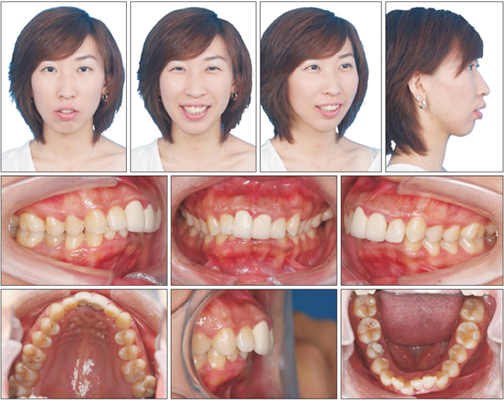
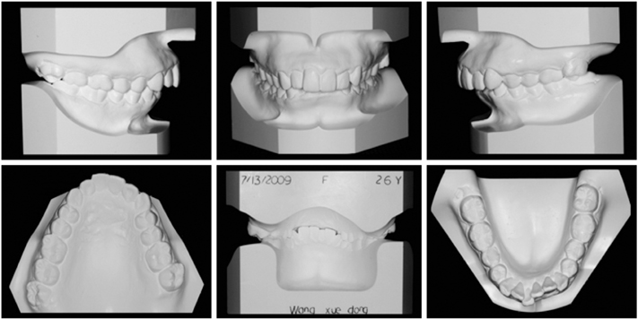
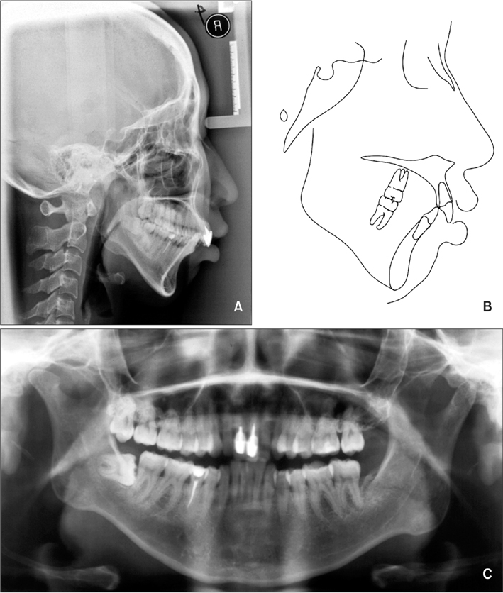
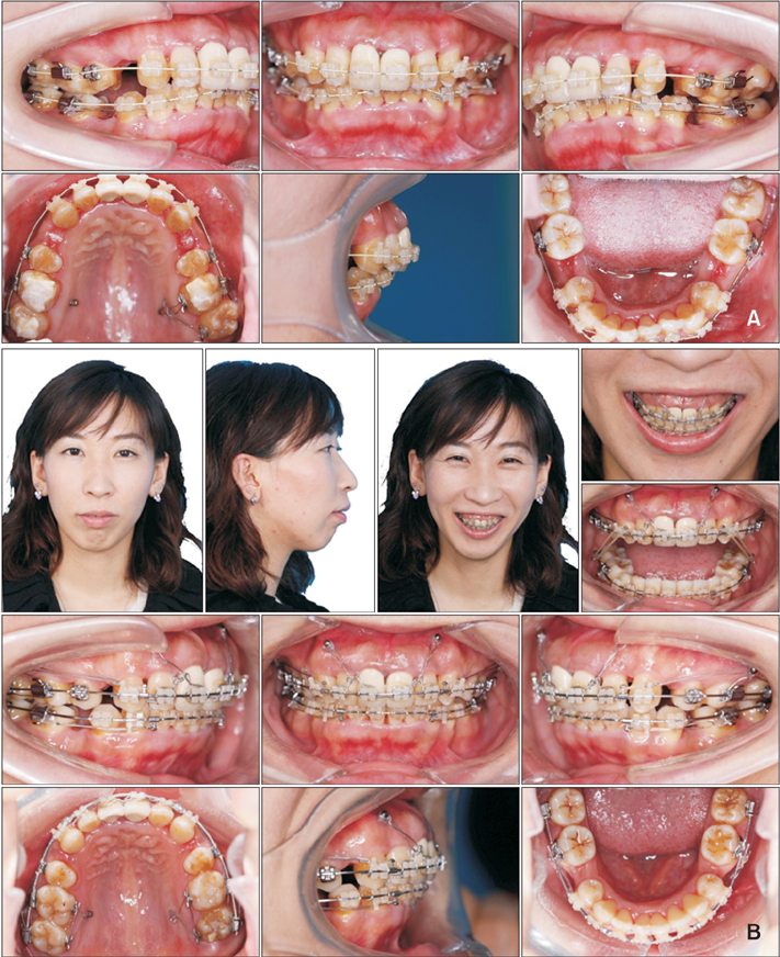
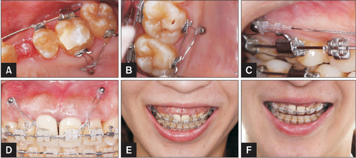
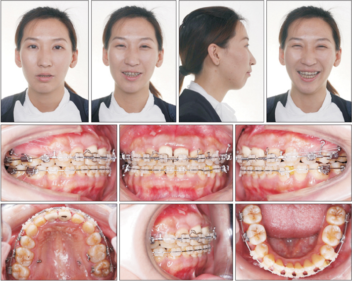
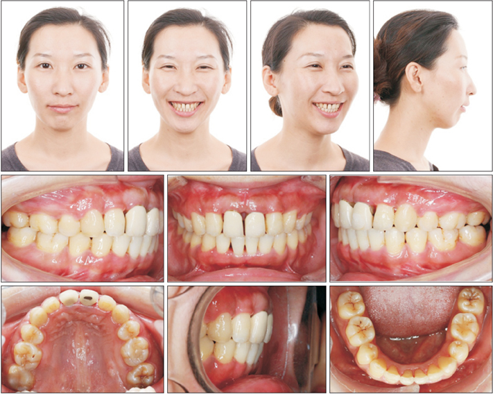
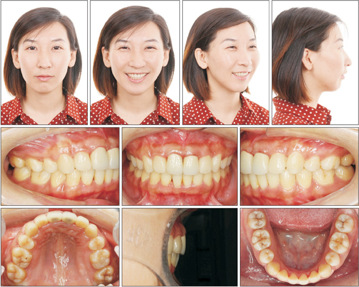
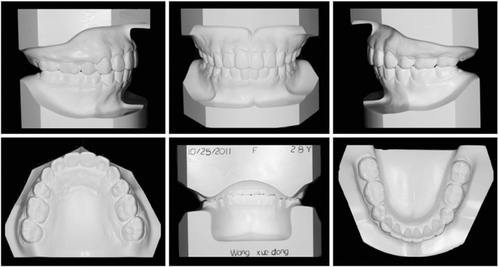
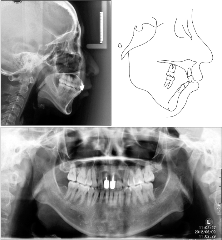
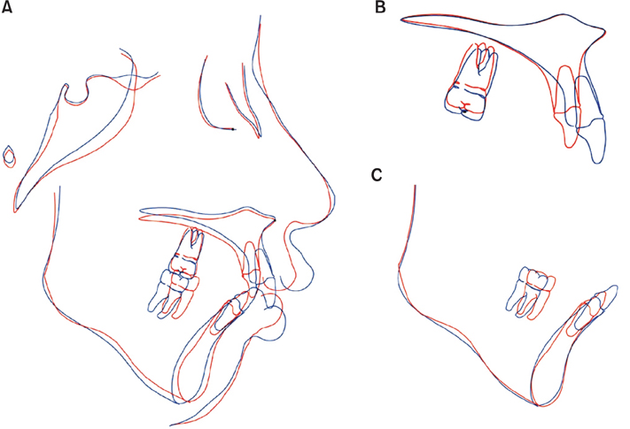
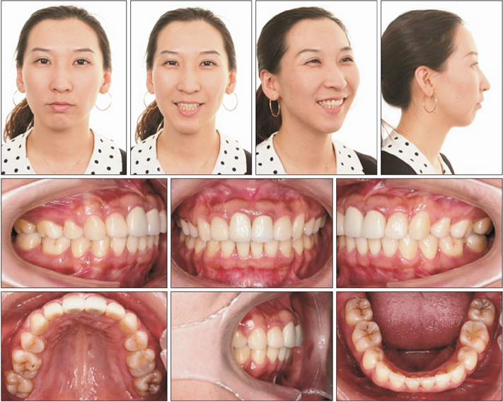
- In the present report, we describe the successful use of miniscrews to achieve vertical control in combination with the conventional sliding MBTâ„¢ straight-wire technique for the treatment of a 26-year-old Chinese woman with a very high mandibular plane angle, deep overbite, retrognathic mandible with backward rotation, prognathic maxilla, and gummy smile. The patient exhibited skeletal Class II malocclusion. Orthodontic miniscrews were placed in the maxillary anterior and posterior segments to provide rigid anchorage and vertical control through intrusion of the incisors and molars. Intrusion and torque control of the maxillary incisors relieved the deep overbite and corrected the gummy smile, while intrusion of the maxillary molars aided in counterclockwise rotation of the mandibular plane, which consequently resulted in an improved facial profile. After 3.5 years of retention, we observed a stable, well-aligned dentition with ideal intercuspation and more harmonious facial contours. Thus, we were able to achieve a satisfactory occlusion, a significantly improved facial profile, and an attractive smile for this patient. The findings from this case suggest that nonsurgical correction using miniscrew anchorage is an effective approach for camouflage treatment of high-angle cases with skeletal Class II malocclusion.
MeSH Terms
Figure
Cited by 1 articles
-
Combined anterior and posterior miniscrews increase apical root resorption of maxillary incisors in protrusion and premolar extraction cases
Zhizun Wang, Li Mei, Zhenxing Tang, Dong Wu, Yue Zhou, Ehab A. Abdulghani, Yuan Li, Wei Zheng, Yu Li
Korean J Orthod. 2025;55(1):26-36. doi: 10.4041/kjod24.136.
Reference
-
1. Canavarro C, Zanella E, Medeiros PJ, Capelli J Jr. Surgical-orthodontic treatment of Class II malocclusion with maxillary vertical excess. J Clin Orthod. 2009; 43:387–392.2. Shu R, Huang L, Bai D. Adult Class II Division 1 patient with severe gummy smile treated with temporary anchorage devices. Am J Orthod Dentofacial Orthop. 2011; 140:97–105.
Article3. Abraham J, Bagchi P, Gupta S, Gupta H, Autar R. Combined orthodontic and surgical correction of adult skeletal class II with hyperdivergent jaws. Natl J Maxillofac Surg. 2012; 3:65–69.
Article4. Bell WH, Jacobs JD, Legan HL. Treatment of Class II deep bite by orthodontic and surgical means. Am J Orthod. 1984; 85:1–20.
Article5. Fowler P. Orthodontics and orthognathic surgery in the combined treatment of an excessively "gummy smile". N Z Dent J. 1999; 95:53–54.6. Nishimura M, Sannohe M, Nagasaka H, Igarashi K, Sugawara J. Nonextraction treatment with temporary skeletal anchorage devices to correct a Class II Division 2 malocclusion with excessive gingival display. Am J Orthod Dentofacial Orthop. 2014; 145:85–94.
Article7. Shimo T, Nishiyama A, Jinno T, Sasaki A. Severe gummy smile with class II malocclusion treated with LeFort I osteotomy combined with horseshoe osteotomy and intraoral vertical ramus osteotomy. Acta Med Okayama. 2013; 67:55–60.8. Upadhyay M, Yadav S, Nanda R. Vertical-dimension control during en-masse retraction with mini-implant anchorage. Am J Orthod Dentofacial Orthop. 2010; 138:96–108.
Article9. Hong RK, Lim SM, Heo JM, Baek SH. Orthodontic treatment of gummy smile by maxillary total intrusion with a midpalatal absolute anchorage system. Korean J Orthod. 2013; 43:147–158.
Article10. Cacciafesta V, Bumann A, Cho HJ, Graham JW, Paquette DE, Park DE, et al. Skeletal anchorage, part 1. J Clin Orthod. 2009; 43:303–317.11. Jung MH. Treatment of severe scissor bite in a middle-aged adult patient with orthodontic mini-implants. Am J Orthod Dentofacial Orthop. 2011; 139:4 Suppl. S154–S165.
Article12. Lin JC, Liou EJ, Bowman SJ. Simultaneous reduction in vertical dimension and gummy smile using miniscrew anchorage. J Clin Orthod. 2010; 44:157–170.13. Ouyang L, Zhou YH, Fu MK, Ding P. Extraction treatment of an adult patient with severe bimaxillary dentoalveolar protrusion using microscrew anchorage. Chin Med J (Engl). 2007; 120:1732–1736.
Article14. Redlich M, Mazor Z, Brezniak N. Severe high Angle Class II Division 1 malocclusion with vertical maxillary excess and gummy smile: a case report. Am J Orthod Dentofacial Orthop. 1999; 116:317–320.
Article15. Kaku M, Kojima S, Sumi H, Koseki H, Abedini S, Motokawa H, et al. Gummy smile and facial profile correction using miniscrew anchorage. Angle Orthod. 2012; 82:170–177.
Article16. Hans MG, Groisser G, Damon C, Amberman D, Nelson S, Palomo JM. Cephalometric changes in overbite and vertical facial height after removal of 4 first molars or first premolars. Am J Orthod Dentofacial Orthop. 2006; 130:183–188.
Article17. Wang Y, Yu H, Jiang C, Li J, An S, Chen Q, et al. Vertical changes in Class I malocclusion between 2 different extraction patterns. Saudi Med J. 2013; 34:302–306.18. Aras A. Vertical changes following orthodontic extraction treatment in skeletal open bite subjects. Eur J Orthod. 2002; 24:407–416.
Article19. Sivakumar A, Valiathan A. Cephalometric assessment of dentofacial vertical changes in Class I subjects treated with and without extraction. Am J Orthod Dentofacial Orthop. 2008; 133:869–875.
Article20. Gkantidis N, Halazonetis DJ, Alexandropoulos E, Haralabakis NB. Treatment strategies for patients with hyperdivergent Class II Division 1 malocclusion: is vertical dimension affected? Am J Orthod Dentofacial Orthop. 2011; 140:346–355.
Article21. Hart TR, Cousley RR, Fishman LS, Tallents RH. Dentoskeletal changes following mini-implant molar intrusion in anterior open bite patients. Angle Orthod. 2015; 85:941–948.
Article22. Kim TW, Kim H, Lee SJ. Correction of deep overbite and gummy smile by using a mini-implant with a segmented wire in a growing class II division 2 patient. Am J Orthod Dentofacial Orthop. 2006; 130:676–685.
Article23. Polat-Ozsoy O, Arman-Ozcirpici A, Veziroglu F. Miniscrews for upper incisor intrusion. Eur J Orthod. 2009; 31:412–416.
Article24. Kalia A, Sharif K. Scissor-bite correction using miniscrew anchorage. J Clin Orthod. 2012; 46:573–579. quiz 583.25. Wilson GB, Kidner G, Hemmings K. The use of implants at an increased vertical dimension of occlusion to correct a scissor bite: a case report. Eur J Prosthodont Restor Dent. 2008; 16:67–72.26. Alani A, Bishop K, Knox J, Gravenor C. The use of implants for anchorage in the correction of unilateral crossbites. Eur J Prosthodont Restor Dent. 2010; 18:123–127.
- Full Text Links
- Actions
-
Cited
- CITED
-
- Close
- Share
- Similar articles
-
- A study of deep overbite and open bite by vertical cephalometric analysis
- A cephalometric study on mesiodistal axial inclination of posterior teeth in open bite and deep bite
- Treatment and posttreatment changes following intrusion of maxillary posterior teeth with miniscrew implants for open bite correction
- A roentgenocephalometric study on the skeletal factors in open-bite and deep-bite
- Nonsurgical maxillary expansion in a 60-year-old patient with gingival recession and crowding

