The relationships between the arrangement of teeth, root resorption, and dental maturity in bovine mandibular incisors
- Affiliations
-
- 1Department of Orthodontic Science, Division of Oral Health Sciences, Graduate School of Medical and Dental Sciences, Tokyo Medical and Dental University, Tokyo, Japan. y.matsumoto.orts@tmd.ac.jp
- KMID: 2392183
- DOI: http://doi.org/10.4041/kjod.2017.47.6.365
Abstract
OBJECTIVE
The objective of this study is to investigate the eruption pattern and root resorption of the bovine anterior dentition in relation to growth-related parameters based on dental maturity.
METHODS
A cross-sectional study was conducted on 110 bovine anterior mandibles by using standard radiography, cone-beam computed tomography (CBCT), and actual measurements. We determined the relationships between the stages of dental maturity by using a modification of Demirjian's method and various growth-related parameters, such as the activity of the root-resorbing tissue and mobility of the deciduous teeth. The correlation of growth-related parameters with interdental spacing and distal unusual root resorption (DRR) of the deciduous fourth incisor was assessed. The cause of mesial unusual root resorption (MRR) of the deciduous fourth incisor was determined on the basis of the arrangement of the permanent third incisor.
RESULTS
An independent t-test and chi-square test indicated significant differences in growth-related parameters associated with dental arch length discrepancy and factors related to the shedding of deciduous teeth between the low and high dental maturity groups. The samples with interdental spacing and DRR showed a larger sum of mesiodistal permanent crown widths and higher dental maturity than did the respective controls. Samples with MRR tended to show a lingually rotated distal tip of the adjacent tooth crown.
CONCLUSIONS
Dental maturity has relevance to the interdental spaces and unusual root resorption of mixed dentition. The position of the adjacent tooth crown on CBCT may be correlated with the occurrence of unusual root resorption of the incisor.
MeSH Terms
Figure
-
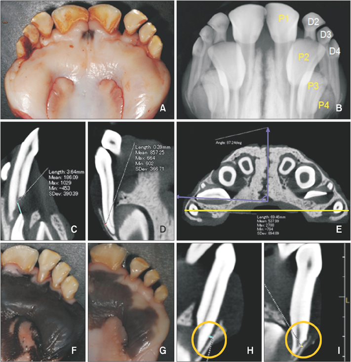
Figure 1 Anterior deciduous and permanent teeth of the cattle, measurements on cone-beam computed tomographic images, classification with respect to spaced dentition, and distal unusual root resorption of the fourth deciduous incisor root (DRR). A, Lingual view; B, standard radiographic image in the same position; C, section in the sagittal direction; D, section in the coronal direction; E, section in the horizontal direction; F, non-spacing group; G, spacing group; H, non-DRR group; I, DRR group. In the sagittal direction, from the plane perpendicular to the Frankfurt plane, the shortest distance between the root surface of D3 and crown of P3 is measured (C). In the coronal direction, from the plane perpendicular to the Frankfurt plane, the narrowest thickness of the periodontal ligament space in the D4 distal root surface is measured (D). In the horizontal directions, in the area parallel to the Frankfurt plane, the longest mandibular bone width and angulation of the P3 crown are measured (E). Black line, mandibular bone width; vertical white line, median suture line; horizontal white line, mesio-distal line of the P3 crown; white angle, angulation of the P3 crown (E). Based on the aspect of spaced dentition, the samples are classified into two groups. The non-spacing group (F) shows no interdental spaces in the deciduous dentition. The spacing group (G) has interdental spaces between the deciduous teeth. Based on the occurrence of DRR, the samples are classified into two groups: non-DRR (H) and DRR groups (I). DRR occurs at the site wherein the distal root surface of D4 is in contact with the alveolar bone. P1, First anterior permanent incisor; P2, second permanent incisor; P3, third anterior permanent incisor; P4, fourth anterior permanent incisor; D2, second deciduous incisor; D3, third deciduous incisor; D4, fourth deciduous incisor.
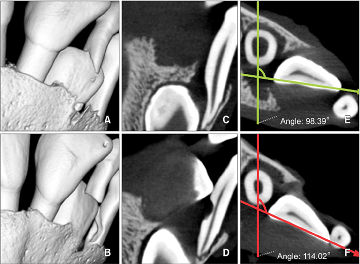
Figure 2 Comparison between the unaffected controls and the samples with mesial unusual root resorption of the fourth deciduous incisors (MRR) by using cone-beam computed tomographic three- and two-dimensional images. The vertical position of the P3 crown and resorption degree of the D3 root are similar in both samples. The distal tip of the P3 crown is located on the labial side with respect to the D4 root in normal samples (A), and the lingual side in MRR samples (B). The contact between the distal tip of the P3 crown and the D4 root surface occurs only in MRR samples in the coronal section (C, D). Angulation of the P3 crown is higher in the samples with MRR (F) than in the normal samples (E) in the horizontal section. Horizontal line in (E) and (F), mesio-distal line of the P3 crown; vertical line in (E) and (F), a line parallel to the median suture line.
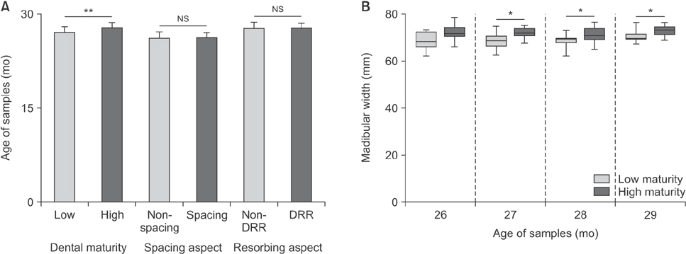
Figure 3 Comparison of chronological age based on dental maturity, spacing aspect, and root-resorbing aspect (A), and comparison of mandibular bone width in the same age range (B). A significant difference between the low and high dental maturity groups is observed (low maturity, 27.3 ± 1.3 months; high maturity, 28.3 ± 1.1 months). However, no significant difference (NS) is observed between the spacing and non-spacing groups (non-spacing, 26.5 ± 1.3 months; spacing, 26.7 ± 1.3 months), and between the distal unusual root resorption (DRR) group and the non-DRR group (non- DRR, 27.8 ± 1.4 months; DRR, 28.1 ± 1.0 months). In the same age range, the mandibular width is higher in the highmaturity than low-maturity samples. The samples with high maturity show a tendency towards a high skeletal growth status (*p < 0.05, **p < 0.001).
Reference
-
1. Hixon EH, Oldfather RE. Estimation of the sizes of unerupted cuspid and bicuspid teeth. Angle Orthod. 1958; 28:236–240.2. Bagherpour A, Pousti M, Adelianfar E. Hand skeletal maturity and its correlation with mandibular dental development. J Clin Exp Dent. 2014; 6:e275–e279.
Article3. Muller-Bolla M, Lupi-Pégurier L, Quatrehomme G, Velly AM, Bolla M. Age estimation from teeth in children and adolescents. J Forensic Sci. 2003; 48:140–148.
Article4. Green LJ. The interrelationships among height, weight and chronological, dental and skeletal ages. Angle Orthod. 1961; 31:189–193.5. Palanisamy V, Rao A, Shenoy R, Baranya SS. Correlation of dental age, skeletal age, and chronological age among children aged 9-14 years: a retrospective study. J Indian Soc Pedod Prev Dent. 2016; 34:310–314.
Article6. Panchbhai AS. Dental radiographic indicators, a key to age estimation. Dentomaxillofac Radiol. 2011; 40:199–212.
Article7. Demirjian A, Goldstein H, Tanner JM. A new system of dental age assessment. Hum Biol. 1973; 45:211–227.8. Bishara SE, Khadivi P, Jakobsen JR. Changes in tooth size-arch length relationships from the deciduous to the permanent dentition: a longitudinal study. Am J Orthod Dentofacial Orthop. 1995; 108:607–613.
Article9. Bishara SE, Jakobsen JR. Individual variation in tooth-size/ arch-length changes from the primary to permanent dentitions. World J Orthod. 2006; 7:145–153.10. el-Nofely A, Sadek L, Soliman N. Spacing in the human deciduous dentition in relation to tooth size and dental arch size. Arch Oral Biol. 1989; 34:437–441.
Article11. Ericson S, Kurol J. Resorption of maxillary lateral incisors caused by ectopic eruption of the canines. A clinical and radiographic analysis of predisposing factors. Am J Orthod Dentofacial Orthop. 1988; 94:503–513.
Article12. Rozylo-Kalinowska I, Kolasa-Raczka A, Kalinowski P. Dental age in patients with impacted maxillary canines related to the position of the impacted teeth. Eur J Orthod. 2011; 33:492–497.
Article13. Dalessandri D, Parrini S, Rubiano R, Gallone D, Migliorati M. Impacted and transmigrant mandibular canines incidence, aetiology, and treatment: a systematic review. Eur J Orthod. 2017; 39:161–169.
Article14. Saka H, Koyama T, Tamatsu Y, Usami A, Ide Y. The morphological studies of root resorption of maxillary primary canines and their relation with the position of successive permanent teeth using Micro-CT. Pediatr Dent J. 2011; 21:145–153.
Article15. Oguchi H. In vitro studies of bone resorption by the root-resorbing tissue from the bovine deciduous tooth. Bull Tokyo Med Dent Univ. 1975; 22:175–183.16. Lind V. Short root anomaly. Scand J Dent Res. 1972; 80:85–93.
Article17. Kumar CL, Sridhar MS. Estimation of the age of an individual based on times of eruption of permanent teeth. Forensic Sci Int. 1990; 48:1–7.
Article18. Andrews AH. Eruption of the maxillary first and second molar teeth in cattle as a method of estimating age. Br Vet J. 1983; 139:355–360.
Article19. Brown WAB, Chapman NG. Age assessment of red deer (Cervus elaphus): from a scoring scheme based on radiographs of developing permanent molariform teeth. J Zoology. 1991; 225:85–97.
Article20. Brown WA, Christofferson PV, Massler M, Weiss MB. Postnatal tooth development in cattle. Am J Vet Res. 1960; 21:7–34.21. Boulanger G. La Calcification des premolaires et molaires et ses relations avecl'Page chronologique et squellettique chez les enfants de 6a 11 ans. Inaugural dissertation. University of Zurich;1958.22. Barrow GV, White JR. Developmental changes of the maxillary and mandibular dental arches. Angle Orthod. 1952; 22:41–46.23. Nanda RS. Root resorption of deciduous teeth in Indian children. Arch Oral Biol. 1969; 14:1021–1030.
Article24. Sriram CH, Priya VK, Sivakumar N, Reddy KR, Babu PJ, Reddy P. Occlusion of primary dentition in preschool children of Chennai and Hyderabad: A comparative study. Contemp Clin Dent. 2012; 3:31–37.
Article25. Vegesna M, Chandrasekhar R, Chandrappa V. Occlusal characteristics and spacing in primary dentition: a gender comparative cross-sectional study. Int Sch Res Notices. 2014; 2014:512680.
Article26. Krishnan V, Davidovitch Z. Chapter 4 gene and tooth movement. In : Krishnan V, Davidovitch Z, editors. Biological mechanisms of tooth movement. New Jersey: Wiley-Blackwell;2009. p. 56–57.27. Ericson S, Bjerklin K, Falahat B. Does the canine dental follicle ause resorption of permanent incisor roots? A computed tomographic study of erupting maxillary canines. Angle Orthod. 2002; 72:95–104.28. Schindel RH, Sheinis MR. Prediction of maxillary lateral-incisor root resorption using sector analysis of potentially impacted canines. J Clin Orthod. 2013; 47:490–493.29. Guarnieri R, Cavallini C, Vernucci R, Vichi M, Leonardi R, Barbato E. Impacted maxillary canines and root resorption of adjacent teeth: a retrospective observational study. Med Oral Patol Oral Cir Bucal. 2016; 21:e743–e750.
Article
- Full Text Links
-
- Actions
-
Cited
- CITED
-
- Close
- Share
-
- Similar articles
-
- A radiographic study on root resorption in the malocclusion patients before orthodontic treatment
- A roentgenographic study on apical root resorption of human permanent teeth
- Pressure Root Resorption of the Second Molar Caused by Third Molar Impaction: A Case Report of Severely Resorbed Root with Vital Pulp
- Factors affecting orthodontically induced root resorption of maxillary central incisors in the Korean population
- Retrospective Analysis of Incisor Root Resorption Associated with Impacted Maxillary Canines
