An Unusual Imaging Finding of Breast Metastasis from Rhabdomyosarcoma
- Affiliations
-
- 1Department of Radiology, College of Medicine, Ewha Womans University Mokdong Hospital, Seoul, Korea. escha@ewha.ac.kr
- KMID: 2392120
- DOI: http://doi.org/10.3348/jksr.2017.77.4.237
Abstract
- Rhabdomyosarcoma mainly occurs in the pediatric age group, with the primary tumor originating from the trunk, neck, and extremities. Metastasis of rhabdomyosarcoma to the breast is very rare. Previous reports have suggested that the mammographic finding of breast rhabdomyosarcoma is an oval-shaped mass with irregular margins and the US finding is a solitary nodular lesion. We report a case of breast metastasis from pleural rhabdomyosarcoma in a 21-year-old woman, presenting as diffuse non-mass involvement and edematous change without a nodular mass.
MeSH Terms
Figure
-
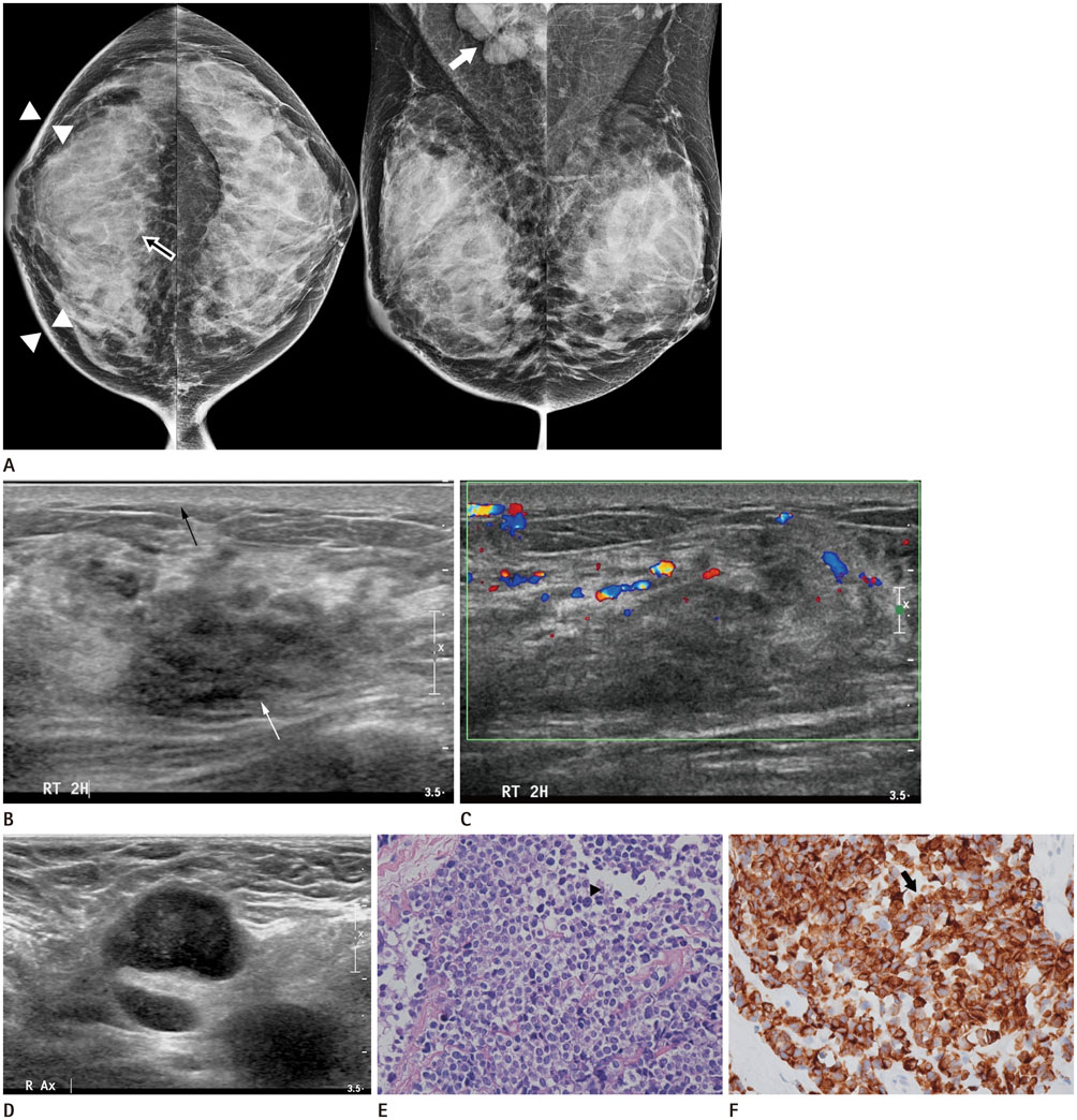
Fig. 1 Breast metastasis from rhabdomyosarcoma in a 21-year-old female. A. Mammogram reveals global asymmetry (empty arrow) with diffuse skin thickening (arrowheads) in the right breast and several enlarged lymph nodes in the right axilla (white arrow). B, C. US reveals an ill-defined hypoechoic lesion (white arrow) with overlying skin thickening (black arrow) in the palpable site of the right breast. Increased vascularity around the lesion is demonstrated. D. There are a few enlarged lymph nodes with loss of fatty hilum in the right axilla. E. Microscopic section of the right breast mass. It shows diffuse growth pattern with small round cell composition. Tumor cells have hyperchromatic nuclei and abundant cytoplasm (arrowhead) (hematoxylin-eosin stain, × 400). F. Tumor cells immunostained for desmin shows a strong reaction (arrow).
Reference
-
1. Jung SP, Lee Y, Han KM, Lee SK, Kim S, Bae SY, et al. Breast metastasis from rhabdomyosarcoma of the anus in an adolescent female. J Breast Cancer. 2013; 16:345–348.2. D'Angelo P, Carli M, Ferrari A, Manzitti C, Mura R, Miglionico L, et al. Breast metastases in children and adolescents with rhabdomyosarcoma: experience of the Italian Soft Tissue Sarcoma Committee. Pediatr Blood Cancer. 2010; 55:1306–1309.3. Mun SH, Ko EY, Han BK, Shin JH, Kim SJ, Cho EY. Breast metastases from extramammary malignancies: typical and atypical ultrasound features. Korean J Radiol. 2014; 15:20–28.4. Birjawi GA, Haddad MC, Tawil AN, Khoury NJ. Metastatic rhabdomyosarcoma to the breast. Eur Radiol. 2001; 11:555–558.5. Perlet C, Sittek H, Forstpointner R, Kessler M, Reiser M. Metastases to the breast from rhabdomyosarcoma: appearances on MRI. Eur Radiol. 1999; 9:1113–1116.6. Yang WT, Kwan WH, Chow LT, Metreweli C. Unusual sonographic appearance with color Doppler imaging of bilateral breast metastases in a patient with alveolar rhabdomyosarcoma of an extremity. J Ultrasound Med. 1996; 15:531–533.7. Binokay F, Soyupak SK, Inal M, Celiktas M, Akgül E, Aksungur E. Primary and metastatic rhabdomyosarcoma in the breast: report of two pediatric cases. Eur J Radiol. 2003; 48:282–284.8. Ahn SJ, Kim SK, Kim EK. Metastatic breast cancer from rhabdomyosarcoma mimicking normal breast parenchyma on sonography. J Ultrasound Med. 2010; 29:489–492.9. Yang WT, Metreweli C. Sonography of nonmammary malignancies of the breast. AJR Am J Roentgenol. 1999; 172:343–348.10. Sheen-Chen SM, Eng HL, Ko SF. Metastatic rhabdomyosarcoma to the breast. Anticancer Res. 2005; 25:527–529.
- Full Text Links
-
- Actions
-
Cited
- CITED
-
- Close
- Share
-
- Similar articles
-
- Metastasis of Rhabdomyosarcoma to the Male Breast: a Case Report with Magnetic Resonance Imaging Findings
- Giant Breast Involvement in Acute Lymphoblastic Leukemia: MRI Findings
- Sinonasal Rhabdomyosarcoma Metastasis in Bilateral Multiple Extraocular Muscles: A Case Report and Brief Literature Review
- A Case of Paratesticular Rhabdomyosarcoma
- A Case of Rhabdomyosarcoma of the Bladder in an Old Adult
