J Rheum Dis.
2011 Jun;18(2):137-141.
Dual-Energy CT as a New Diagnostic Tool for Gout
- Affiliations
-
- 1Department of Internal Medicine, Wonkwang University College of Medicine, Iksan, Korea.
- 2Department of Radiology, Wonkwang University College of Medicine, Iksan, Korea. juhngsk@wonkwang.ac.kr
Abstract
- Gout is the most common crystal-associated arthropathy. Gout is caused by deposition of monosodium urate crystals within the joints, and it is often associated with hyperuricemia. Acute gout involves the first metatarsophalangeal joint (podagra) in approximately 50% of cases and its peak incidence occurs in middle age. Although the clinical features can help with making the diagnosis of gout, many inflammatory diseases such as cellulitis, pseudogout and septic arthritis can mimic or coexist with it. The definitive diagnosis requires polarized light microscopy of the fluid aspirated from the involved joint and this shows needle-shaped, negative birefringent monosodium urate crystals. However, joint aspiration can be technically difficult, and none of the conventional imaging modalities for gout specifically identifies the chemical composition of uric acid. The advent of Dual-Energy CT (DECT) is a noninvasive method that has the potential to confirm gout and monitor the response to treatment. DECT scan can show monosodium urate deposition by using color coding. The authors performed DECT scans for detecting uric acid deposition and confirming the gout noninvasively.
Keyword
MeSH Terms
Figure
-
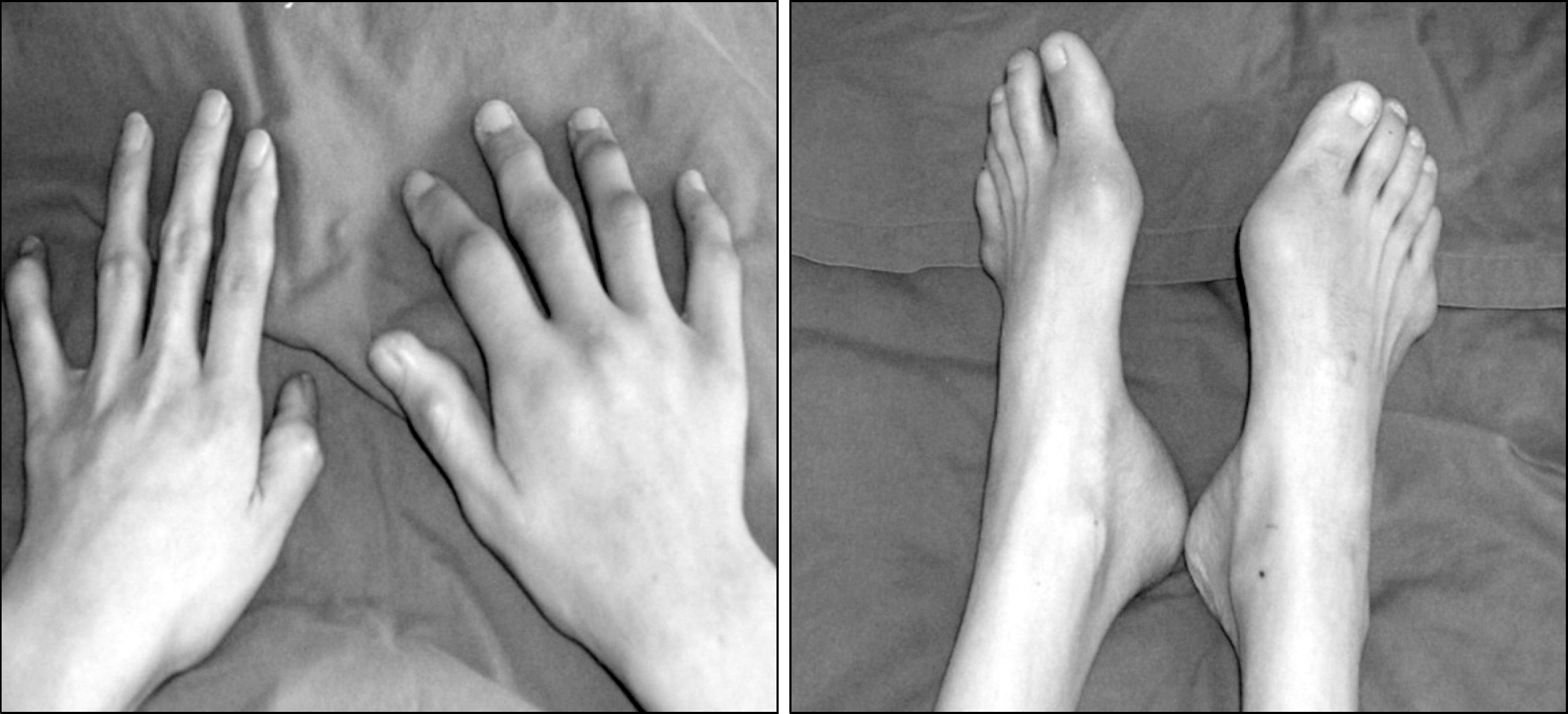
Figure 1. The images visualize multiple tophi around the proximal interphalangeal joints of the right hand and the 1 st metatarsophalang-eal joint of the left foot.
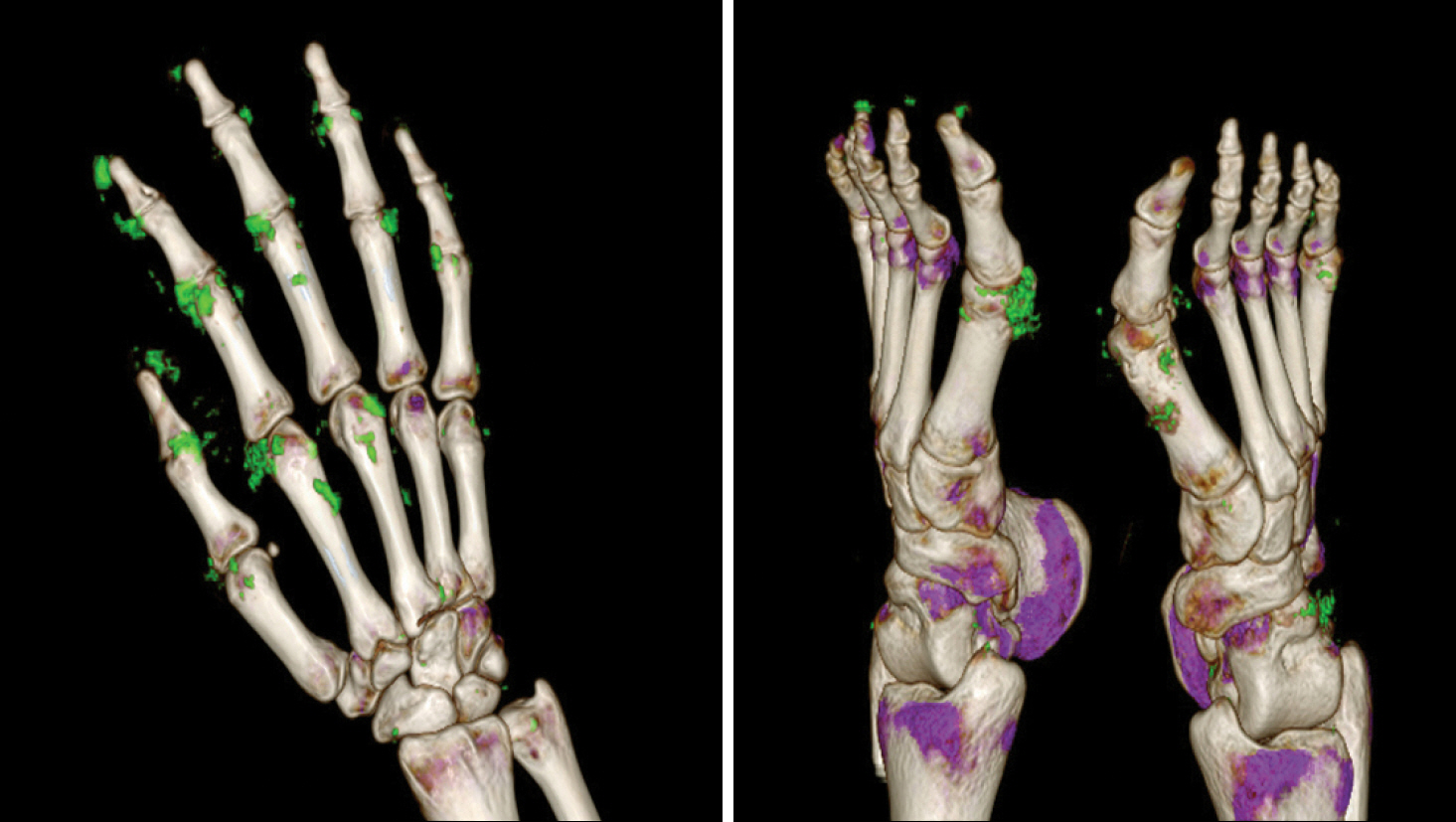
Figure 2. The dual energy computed tomography images demons-trate uric acid deposits (green color) on multiple joints of the right hand and both feet.
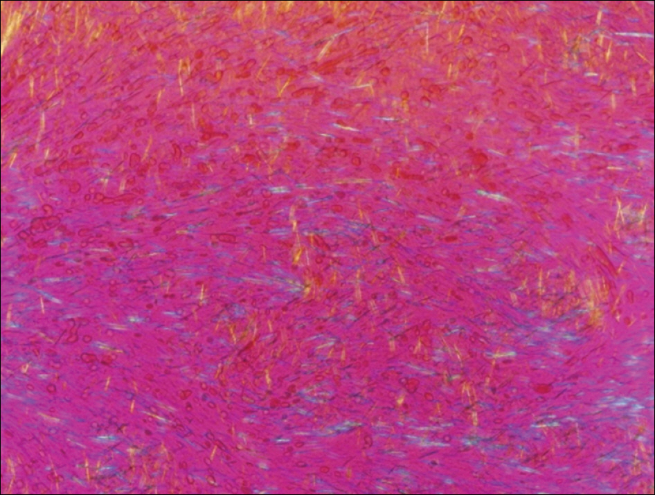
Figure 3. Repolarizing microscopy of the aspirated fluid from the left knee shows negative birefringent needle-shaped crystals, indica-ting monosodium urate crystals.
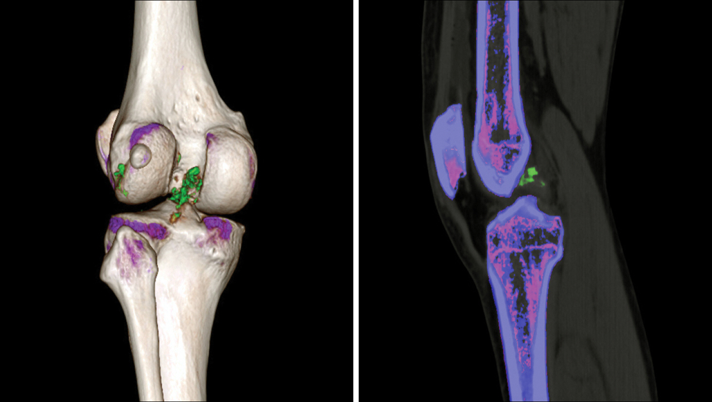
Figure 4. The dual energy computed tomography images demonst-rate uric acid deposits (green color) on the posterior cruciate ligament of the left knee.
Reference
-
References
1. Choi IY, Whang KS, Ahn SC, Kim YH. A clinical study on gouty arthritis. J Korean Rheum Assoc. 1994; 1:169–74.2. Choi HK, Mount DB, Reginato AM. American College of Physicians; American Physiological Society. Pathogenesis of gout. Ann Intern Med. 2005; 143:499–516.
Article3. Monu JU, Pope TL Jr. Gout: a clinical and radiologic review. Radiol Clin North Am. 2004; 42:169–84.
Article4. Thiele RG, Schlesinger N. Diagnosis of gout by ultra-sound. Rheumatology (Oxford). 2007; 46:1116–21.
Article5. Schumacher HR Jr, Becker MA, Edwards NL, Palmer WE, MacDonald PA, Palo W, et al. Magnetic resonance imaging in the quantitative assessment of gouty tophi. Int J Clin Pract. 2006; 60:408–14.
Article6. Reimann AJ, Rinck D, Birinci-Aydogan A, Scheuering M, Burgstahler C, Schroeder S, et al. Dual-source computed tomography: advances of improved temporal reso-lution in coronary plaque imaging. Invest Radiol. 2007; 42:196–203.7. Johnson TR, Krauss B, Sedlmair M, Grasruck M, Bruder H, Morhard D, et al. Material differentiation by dual energy CT: initial experience. Eur Radiol. 2007; 17:1510–7.
Article8. Graser A, Johnson TR, Chandarana H, Macari M. Dual energy CT: preliminary observations and potential clinical applications in the abdomen. Eur Radiol. 2009; 19:13–23.
Article9. Nicolaou S, Yong-Hing CJ, Galea-Soler S, Hou DJ, Louis L, Munk P. Dual-energy CT as a potential new diagnostic tool in the management of gout in the acute setting. AJR Am J Roentgenol. 2010; 194:1072–8.
Article10. Choi HK, Al-Arfaj AM, Eftekhari A, Munk PL, Shojania K, Reid G, et al. Dual energy computed tomography in tophaceous gout. Ann Rheum Dis. 2009; 68:1609–12.
Article11. Schumacher HR Jr, Becker MA, Edwards NL, Palmer WE, MacDonald PA, Palo W, et al. Magnetic resonance imaging in the quantitative assessment of gouty tophi. Int J Clin Pract. 2006; 60:408–14.
Article12. Anonymous. Report of the united nations scientific com-mittee on the effects of atomic radiation to the general assembly. 2000.
- Full Text Links
-
- Actions
-
Cited
- CITED
-
- Close
- Share
-
- Similar articles
-
- Dual-Energy CT: New Horizon in Medical Imaging
- Ultrasonography and dual-energy computed tomography: impact for the detection of gouty deposits
- Spectral Computed Tomography: Fundamental Principles and Recent Developments
- Dual-Layer Computed Tomography in Cardiovascular Imaging
- Confirmation of Tophi in a Patient of Gout using Dual-Energy CT
