Mid-foot retinaculum: an unrecognized entity
- Affiliations
-
- 1Department of Anatomy, K. S. Hegde Medical Academy, Deralakatte, Mangalore, India. swathishriya@gmail.com
- 2Department of Anatomy, Academy of Medical Sciences, Pariyaram, Kannur, India.
- 3Department of Anatomy, All India Institute of Medical Sciences, Bhopal, India.
- KMID: 2390477
- DOI: http://doi.org/10.5115/acb.2017.50.3.171
Abstract
- Retinacula are thickenings of deep fascia in the region of joints that hold down the tendons preventing them from bowing out of position. In the region of ankle, number of such retinacula have been described. Retinacula like superior and inferior extensor retinacula have been described which hold down the tendons of leg muscles passing to the foot beneath them. As the extensor tendons of the leg have more distal attachment to the toes, the present study was conducted to ascertain the presence of any additional retinaculum in the mid-foot region, which would tie down the tendons for their effective action at the distal joints. The aim was also to determine the attachments of the retinaculum, if present as well as the structures passing beneath them. Fifty cadaveric feet were dissected carefully for this purpose. Presence of an additional extensor retinaculum distal to the inferior band of inferior extensor retinaculum in the mid-foot region was found in 22 feet. Besides the extensor tendons, medial terminal branch of deep peroneal nerve and dorsalis pedis artery was found to pass beneath the retinaculum. A partial or complete mid-foot retinaculum existed in the mid-foot region covering the tarsometatarsal joints in about half of study population. Functionally, this retinaculum may prevent bowstringing of the extensor tendons, clinically it may predispose to entrapment of deep peroneal nerve mimicking anterior tarsal tunnel syndrome.
Keyword
MeSH Terms
Figure
-
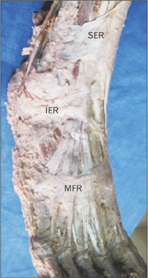
Fig. 1 Complete mid foot retinaculum. Superior extensor retinaculum (SER), inferior extensor retinaculum (IER), and a complete midfoot retinaculum (MFR).

Fig. 2 The medial end of midfoot retinaculum (MFR) blending with plantar fascia (black arrows). Proximally, it is seen merging with the distal limb of inferior extensor retinaculum (IER) and distally it is seen extending up to first metatarsophalangeal joint (I MTP) marked in white arrow.
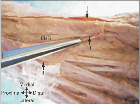
Fig. 3 Different compartments deep to midfoot retinaculum. First, second, and third compartments (I , II, III), and extensor hallucis brevis (EHB).
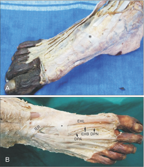
Fig. 4 (A, B) Partial retinaculum (asterisk) in midfoot region. (B) The dorsalis pedis artery (DPA), deep peroneal nerve (DPN), extensor hallucis longus (EHL), extensor hallucis brevis (EHB) deep to the retinaculum. IER, inferior extensor retinaculum.
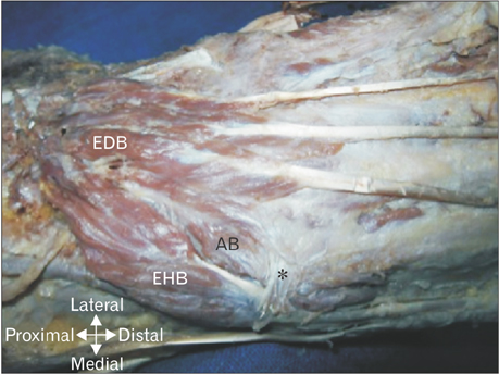
Fig. 5 An additional belly (AB) of extensor digitorum brevis (EDB) passing deep to the retinaculum (asterisk). EHB, extensor hallucis brevis.
Reference
-
1. Muscolino JE. Kinesiology: the skeletal system and muscle function. 2nd ed. St. Louis, MO: Elsevier;2011. p. 306–312.2. Standring S, Ellis H, Healy JC, Johnson D, Williams A, Collins P, Wigley C. Gray's anatomy: the anatomical basis of clinical practice. 39th ed. Edinburgh: Churchill Livingstone;2005. p. 1507.3. Sinnatamby CS. Last's anatomy: regional and applied. 12th ed. Edinburgh: Churchill Livingstone;2011. p. 145.4. Sarrafian SK. Anatomy of foot and ankle: descriptive, topographic, functional. 2nd ed. Philadelphia, PA: J. B. Lippincott Co.;1993. p. 123.5. Aktan Ikiz ZA, Ucerler H, Uygur M. Dimensions of the anterior tarsal tunnel and features of the deep peroneal nerve in relation to clinical application. Surg Radiol Anat. 2007; 29:527–530.6. Lawrence SJ, Botte MJ. The deep peroneal nerve in the foot and ankle: an anatomic study. Foot Ankle Int. 1995; 16:724–728.7. Borges LF, Hallett M, Selkoe DJ, Welch K. The anterior tarsal tunnel syndrome: report of two cases. J Neurosurg. 1981; 54:89–92.8. Kennedy JG, Baxter DE. Nerve disorders in dancers. Clin Sports Med. 2008; 27:329–334.9. Liu Z, Zhou J, Zhao L. Anterior tarsal tunnel syndrome. J Bone Joint Surg Br. 1991; 73:470–473.10. Seçıl Y, Tokuçoğlu F, Beckmann Y, Arici Ş, Eryaşar G. Anterior tarsal tunnel syndrome: electrophysiological and clinical evaluation of five cases. J Neurol Sci. 2012; 29:819–825.11. Dellon AL. Deep peroneal nerve entrapment on the dorsum of the foot. Foot Ankle. 1990; 11:73–80.12. Kanbe K, Kubota H, Shirakura K, Hasegawa A, Udagawa E. Entrapment neuropathy of the deep peroneal nerve associated with the extensor hallucis brevis. J Foot Ankle Surg. 1995; 34:560–562.
- Full Text Links
-
- Actions
-
Cited
- CITED
-
- Close
- Share
-
- Similar articles
-
- Operative Treatment of Chronic Recurrent Dislocation of Peroneal Tendon: A Case Report
- The Checkrein Deformity of Extensor Hallucis Longus Tendon and Extensor Retinaculum Syndrome with Deep Peroneal Nerve Entrapment after Triplane Fracture: A Case Report
- Surgical Treatment of Muller-Weiss Disease: A Case Report
- Peroneus Longus Dislocation associated with Trimalleolar Fracture: A Case Report
- Operative Treatment of Acute Peroneal Tendon Subluxation in Athletes: A Case Report - 2 Cases
