Anat Cell Biol.
2017 Mar;50(1):73-75. 10.5115/acb.2017.50.1.73.
Recognition of a rare intrathoracic rib with computed tomography: a case report
- Affiliations
-
- 1Department of Anatomical Sciences and Biology, Shahid Beheshti University of Medical Science, Tehran, Iran.
- 2Department of Anatomical Science, Tehran Medical Sciences Branch, Islamic Azad University, Tehran, Iran. shabnam.abdi62@yahoo.com
- KMID: 2390429
- DOI: http://doi.org/10.5115/acb.2017.50.1.73
Abstract
- One of the uncommon congenital variations is intrathoracic rib which a normal, a bifid, or an accessory rib lies within the thoracic cavity that is founded accidentally. Clinically, in most cases they are without symptoms; however, it may cause intrathoracic problems therefore it is important for radiologists and physicians to identify to prevent of excessive intervention and treatment during imaging diagnostic techniques of thoracic problems. In this report, we provide the case of a rare presentation of an intrathoracic rib in a 3-year-old boy arising from the inferior portion of a second rib based on findings from computed tomography. To our knowledge, this is only the second reported case of this type of intrathoracic rib that demonstrated with computed tomography.
Figure
-
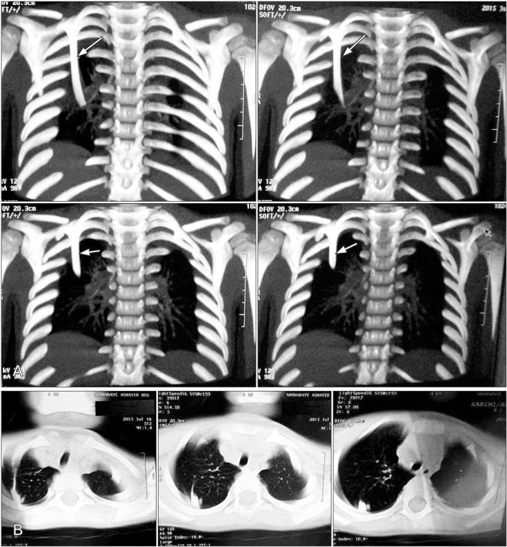
Fig. 1 A 3-year-old boy with rare intrathoracic rib, coronal (A) and axial (B) view of chest computed tomography scan shows arising intrathoracic rib from the inferior border of the right second rib (arrows).
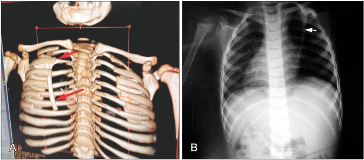
Fig. 2 Volume rendered 3D images (A) and chest radiography (B), a rare intrathoracic originating from the middle part of inferior border of right second rib (ossified, thin, long, and obliquely oriented) (arrows).
Reference
-
1. Mahajan PS, Hasan IA, Ahamad N, Al Moosawi NM. A unique case of left second supernumerary and left third bifid intrathoracic ribs with block vertebrae and hypoplastic left lung. Case Rep Radiol. 2013; 2013:620120. PMID: 24416613.2. Kayiran SM, Gumus T, Kayiran PG, Gurakan B. Supernumerary intrathoracic rib. Arch Dis Child. 2013; 98:441. PMID: 23361140.3. Kamano H, Ishihama T, Ishihama H, Kubota Y, Tanaka T, Satoh K. Bifid intrathoracic rib: a case report and classification of intrathoracic ribs. Intern Med. 2006; 45:627–630. PMID: 16755094.4. Basarslan F, Bayarogullari H, Tutanc M, Arica V, Yilmaz C, Davran R. Intrathoracic rib associated with pulmonary collapse in a pediatric patient. Iran J Radiol. 2012; 9:220–222. PMID: 23408171.5. Chung JH, Pipavath SN. Images in clinical medicine: intrathoracic rib. N Engl J Med. 2009; 361:2557. PMID: 20032325.6. Apaydin M, Sarsilmaz A, Varer M. Third accessory (supernumerary) intrathoracic right rib. Surg Radiol Anat. 2009; 31:641–643. PMID: 19322508.7. Watkins TW, Wilkinson AG, Greer ML. Atypical intrathoracic rib in a pediatric patient requiring helical CT scan with 3-D reconstruction for diagnosis. Pediatr Radiol. 2008; 38:1003–1005. PMID: 18478220.8. Fischer B, Degenhardt P. A complex congenital anomaly: liver eventration because of intrathoracic rib and vertebral segmentation disorder: a case report. J Pediatr Surg. 2008; 43:E5–E7.9. Argyriou P, Pousios D, Tsocanas D, Psathakis C, Panagiotopoulos N, Piyis A. Demonstration of an intrathoracic rib with computed tomography and three-dimensional reconstruction. Ann Thorac Surg. 2007; 84:2117. PMID: 18036957.10. Fainsinger MH. Kohler/Zimmer: borderlands of normal and early pathologic findings in skeletal radiography. 4th English ed. J Bone Joint Surg Am. 1993; 75:1742.11. Lutz BS, Matejic B, Ingianni G. Thoracic outlet syndrome: follow-up on 33 cases with regard to vascular compression. Int J Angiol. 1998; 7:202–205. PMID: 9585450.12. Kermond AJ. Supernumerary intrathoracic rib: an easily recognized rare anomaly. Australas Radiol. 1971; 15:131–132. PMID: 5114465.13. Trigaux JP, Sibille Y, Van Beers B. Intrathoracic rib: CT features. J Comput Assist Tomogr. 1990; 14:133–135. PMID: 2298979.14. Peterson MS, Plunkett MB. CT demonstration of an intrathoracic rib. AJR Am J Roentgenol. 1993; 160:895.15. Branch S, Rogers JM, Brownie CF, Chernoff N. Supernumerary lumbar rib: manifestation of basic alteration in embryonic development of ribs. J Appl Toxicol. 1996; 16:115–119. PMID: 8935784.16. Hawass NE, Bahakim H, al-Boukai AA. Intrathoracic fat: a new CT feature of intrathoracic rib: case report. Clin Imaging. 1991; 15:31–34. PMID: 2059886.
- Full Text Links
-
- Actions
-
Cited
- CITED
-
- Close
- Share
-
- Similar articles
-
- Computed tomographic findings of a primary intrathoracic goiter
- Case Report of a Simple Rib Fracture Caused by Coughing
- Primary Intrathoracic Goiter: A case report
- A Case of Intrathoracic Kidney in an Adult
- Hemothorax Caused by Spontaneous Rupture of Hepatocellular Carcinoma in the Pleural Cavity: A Case Report
