Delayed Absorption of Subretinal Fluid after Retinal Reattachment Surgery and Associated Choroidal Features
- Affiliations
-
- 1Department of Ophthalmology, Samsung Medical Center, Sungkyunkwan University School of Medicine, Seoul, Korea. swkang@skku.edu
- KMID: 2390283
- DOI: http://doi.org/10.3341/kjo.2016.0033
Abstract
- PURPOSE
The aim of this study was to investigate the incidence and associated clinical factors of delayed absorption of subretinal fluid (SRF) after surgery for rhegmatogenous retinal detachment.
METHODS
This study involved 36 eyes of 36 consecutive patients who underwent successful surgery for rhegmatogenous retinal detachment. A complete ophthalmologic evaluation, including clinical fundus examination, optical coherence tomography, and indocyanine green angiography, was conducted before and after surgery. Delayed absorption was defined as the presence of residual concave SRF or an SRF bleb at 6 months after surgery. Clinical factors and choroidal features on indocyanine green angiography were compared according to the presence and absence of delayed absorption.
RESULTS
Eighteen of 36 eyes (50%) showed delayed absorption. Macular involvement and worse preoperative visual acuity were significantly related to the presence of delayed absorption (p = 0.001 and p = 0.034, respectively). On indocyanine green angiography, preoperative choroidal vascular hyperpermeability was noted in 70% of eyes with delayed absorption and in 14% of eyes without it (p = 0.010).
CONCLUSIONS
Delayed absorption of SRF after retinal reattachment surgery was not rare, with a 50% of incidence in this study. Macula-off status was significantly related to the incidence of delayed SRF absorption, and choroidal features such as choroidal vascular hyperpermeability might be responsible in part, possibly through the resultant exudative property of choroid.
Keyword
MeSH Terms
Figure
-
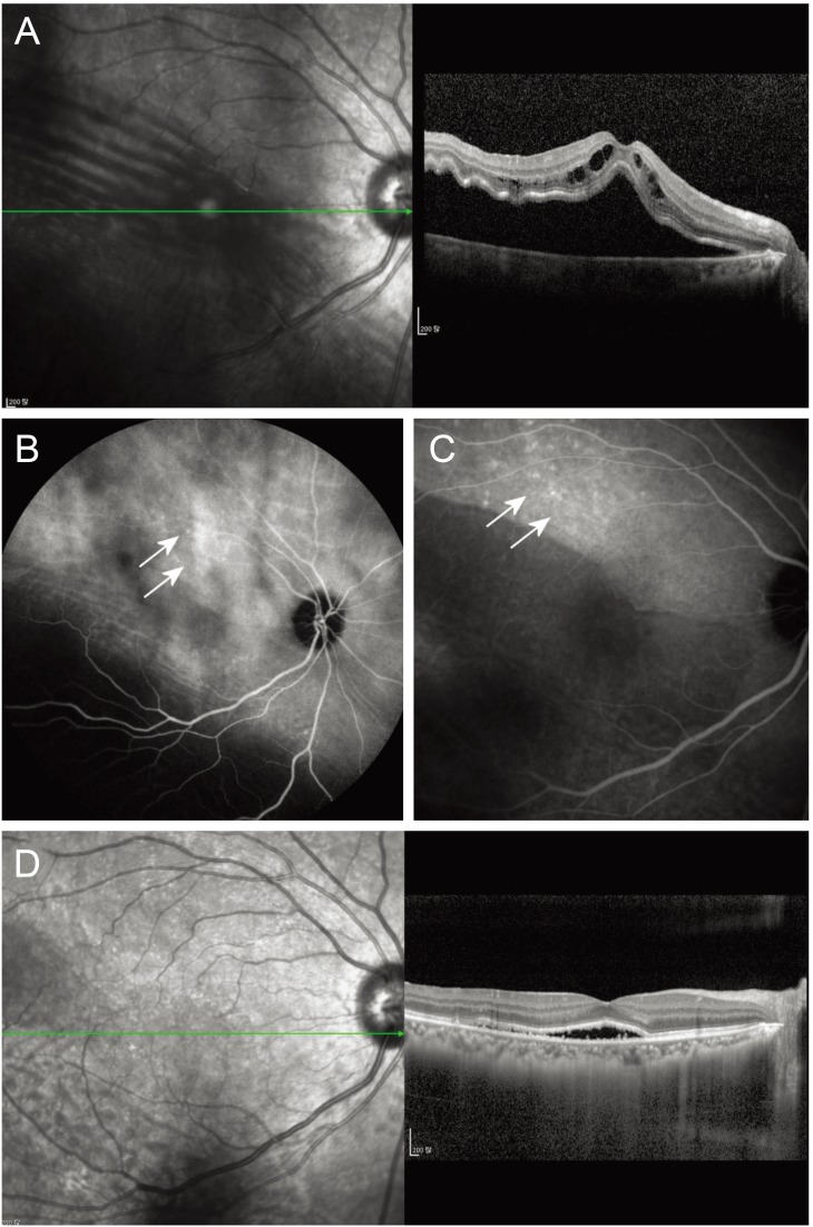
Fig. 1 A case with delayed absorption of subretinal fluid (case 9). (A) Preoperative infrared fundus photography and optical coherence tomography imaging. (B) Preoperative indocyanine green angiography shows choroidal vascular hyperpermeability (arrows) around the macula. (C) Postoperative indocyanine green angiography of the same patient shows areas of hyperfluorescence around subretinal fluid (arrows). (D) A concave subretinal fluid bleb, which progressively decreased in height.
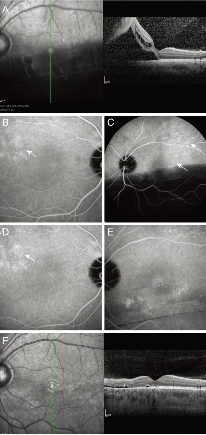
Fig. 2 A case with delayed absorption of subretinal fluid (case 16). (A) Preoperative infrared fundus photography and optical coherence tomography imaging. Preoperative indocyanine green angiography shows focal choroid vascular hyperpermeability (arrow) around the macula in the fellow eye (B) and in the affected eye with retinal detachment (C). (D) Postoperative 1-month indocyanine green angiography shows that preoperatively observed focal hyperfluorescence (arrow) remains in the fellow eye. (E) Patchy hyperfluorescence with choroidal vascular hyperpermeability are observed in the affected eye. (F) Concave subretinal fluid and a subretinal fluid bleb, which progressively decreased in height.
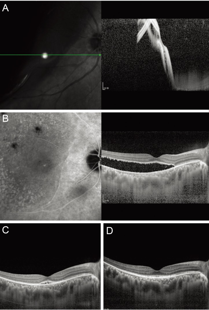
Fig. 3 Multimodal images of a case with persistent subretinal fluid (SRF) where postoperative photodynamic therapy resulted in complete resolution of SRF (case 36). (A) Preoperative infrared fundus photography and optical coherent tomography imaging show macula-off retinal detachment. (B) Indocyanine green angiography and optical coherent tomography taken 1 year postoperatively show concave SRF involving the fovea with thickened choroid. A mild degree of choroidal vascular hyperpermeability is noted from the temporal margin of the optic disk to the fovea. (C) A significant decrease in SRF was observed 3 months after photodynamic therapy. (D) Complete resolution of SRF was confirmed at 6 months after photodynamic therapy.
Reference
-
1. Machemer R. Experimental retinal detachment in the owl monkey. IV. The reattached retina. Am J Ophthalmol. 1968; 66:1075–1091. PMID: 4973290.2. Baba T, Hirose A, Moriyama M, Mochizuki M. Tomographic image and visual recovery of acute macula-off rhegmatogenous retinal detachment. Graefes Arch Clin Exp Ophthalmol. 2004; 242:576–581. PMID: 14997321.
Article3. Benson SE, Schlottmann PG, Bunce C, et al. Optical coherence tomography analysis of the macula after vitrectomy surgery for retinal detachment. Ophthalmology. 2006; 113:1179–1183. PMID: 16647127.
Article4. Hagimura N, Iida T, Suto K, Kishi S. Persistent foveal retinal detachment after successful rhegmatogenous retinal detachment surgery. Am J Ophthalmol. 2002; 133:516–520. PMID: 11931785.
Article5. Kaga T, Fonseca RA, Dantas MA, et al. Optical coherence tomography of bleb-like subretinal lesions after retinal reattachment surgery. Am J Ophthalmol. 2001; 132:120–121. PMID: 11438071.
Article6. Robertson DM. Delayed absorption of subretinal fluid after scleral buckling procedures: the significance of subretinal precipitates. Trans Am Ophthalmol Soc. 1978; 76:557–583. PMID: 754382.7. Wolfensberger TJ, Gonvers M. Optical coherence tomography in the evaluation of incomplete visual acuity recovery after macula-off retinal detachments. Graefes Arch Clin Exp Ophthalmol. 2002; 240:85–89. PMID: 11931084.
Article8. Wolfensberger TJ. Foveal reattachment after macula-off retinal detachment occurs faster after vitrectomy than after buckle surgery. Ophthalmology. 2004; 111:1340–1343. PMID: 15234134.
Article9. Kang SW, Kim JH, Shin WJ, Kim JI. Subretinal fluid bleb after successful scleral buckling and cryotherapy for retinal detachment. Am J Ophthalmol. 2008; 146:205–210. PMID: 18514609.
Article10. Guyer DR, Yannuzzi LA, Slakter JS, et al. Digital indocyanine green videoangiography of central serous chorioretinopathy. Arch Ophthalmol. 1994; 112:1057–1062. PMID: 8053819.
Article11. Chung SE, Kang SW, Lee JH, Kim YT. Choroidal thickness in polypoidal choroidal vasculopathy and exudative age-related macular degeneration. Ophthalmology. 2011; 118:840–845. PMID: 21211846.
Article12. Jirarattanasopa P, Ooto S, Nakata I, et al. Choroidal thickness, vascular hyperpermeability, and complement factor H in age-related macular degeneration and polypoidal choroidal vasculopathy. Invest Ophthalmol Vis Sci. 2012; 53:3663–3672. PMID: 22570352.
Article13. Kim YK, Woo SJ, Kim KE, Park KH. Subretinal fluid resorption. Ophthalmology. 2010; 117:1655. 1655.e1. 1655.e4. PMID: 20682381.
Article14. Kim YK, Woo SJ, Park KH, et al. Comparison of persistent submacular fluid in vitrectomy and scleral buckle surgery for macula-involving retinal detachment. Am J Ophthalmol. 2010; 149:623–629.e1. PMID: 20149338.
Article15. Kim YK, Ahn J, Woo SJ, et al. Multiple subretinal fluid blebs after successful retinal detachment surgery: incidence, risk factors, and presumed pathophysiology. Am J Ophthalmol. 2014; 157:834–841. PMID: 24447856.
Article16. Imamura Y, Engelbert M, Iida T, et al. Polypoidal choroidal vasculopathy: a review. Surv Ophthalmol. 2010; 55:501–515. PMID: 20850857.
Article17. Maruko I, Iida T, Saito M, et al. Clinical characteristics of exudative age-related macular degeneration in Japanese patients. Am J Ophthalmol. 2007; 144:15–22. PMID: 17509509.
Article18. Tsai DC, Chen SJ, Huang CC, et al. Epidemiology of idiopathic central serous chorioretinopathy in Taiwan, 2001-2006: a population-based study. PLoS One. 2013; 8:e66858. PMID: 23826160.
Article19. Iida T, Kishi S, Hagimura N, Shimizu K. Persistent and bilateral choroidal vascular abnormalities in central serous chorioretinopathy. Retina. 1999; 19:508–512. PMID: 10606450.
Article20. Sasahara M, Tsujikawa A, Musashi K, et al. Polypoidal choroidal vasculopathy with choroidal vascular hyperpermeability. Am J Ophthalmol. 2006; 142:601–607. PMID: 17011852.
Article21. Benson SE, Schlottmann PG, Bunce C, et al. Optical coherence tomography analysis of the macula after scleral buckle surgery for retinal detachment. Ophthalmology. 2007; 114:108–112. PMID: 17095091.
Article22. Kim YK, Kim YW, Woo SJ, et al. Persistent submacular fluid and structural and functional recovery of retina. Ophthalmology. 2014; 121:2501–2502. PMID: 25085630.
Article23. Koizumi H, Yamagishi T, Yamazaki T, Kinoshita S. Relationship between clinical characteristics of polypoidal choroidal vasculopathy and choroidal vascular hyperpermeability. Am J Ophthalmol. 2013; 155:305–313.e1. PMID: 23022162.
Article
- Full Text Links
-
- Actions
-
Cited
- CITED
-
- Close
- Share
-
- Similar articles
-
- Delayed Absorption of Subretinal Fluid after Scleral Buckling Procedure for Rhegmatogenous Retinal Detachment
- Drainge vs. Nondrainage of Suvretinal Fluid in Scleral Buckling Procedure
- Pathology of Rhegmatogenous Retinal Detachment
- Clinical Evaluation of Pneumatic Retinopexy
- Clinical Analysis of 137 Eyes of Retinal Detachment Surgery with Drainage of Subretinal Fluid
