Fracture and Dislocation of the Midtarsal Joint: A Case Report
- Affiliations
-
- 1Department of Orthopaedic Surgery, Bundang Jesaeng General Hospital, Daejin Medical Center, Seongnam, Korea. bluejun0929@gmail.com
- KMID: 2389941
- DOI: http://doi.org/10.14193/jkfas.2017.21.3.108
Abstract
- The midtarsal joint is composed of the talonavicular and calcaneocuboid joints. It is also known as the Chopart joint. Midtarsal joint fracture and dislocation are relatively rare and frequently missed or misdiagnosed. A proper understanding about the anatomy of the midtarsal joint is an essential part in comprehending the mechanism of injury and rationale for treatment. Anatomical reduction of midtarsal joint with correction of the column in length and shape are important; however, it is technically challenging and may require open procedure. Herein, we described a case of initial open reduction and internal fixation for midtarsal joint fracture and dislocation with a brief literature review.
Keyword
MeSH Terms
Figure
-
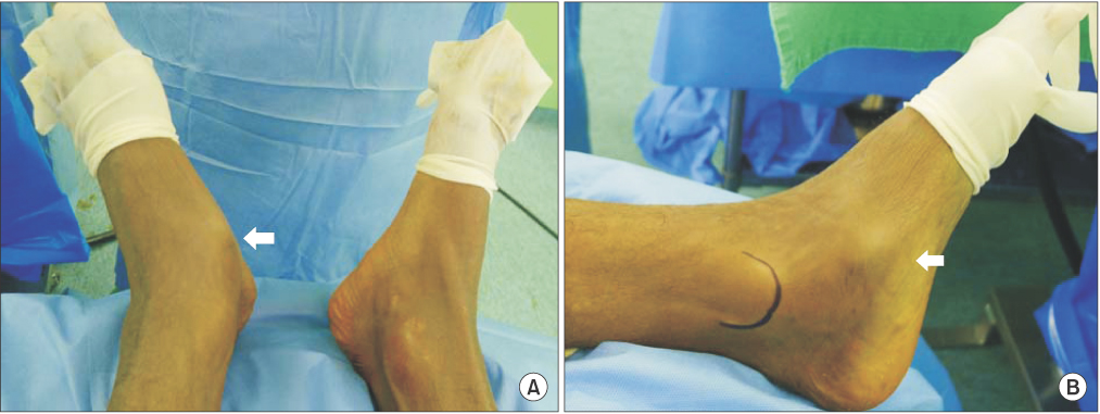
Figure 1 (A, B) Skin tenting (white arrows) is found in medial column because of inferolateral displacement of cuboid bone. Also, the midfoot and forefoot of left foot are more lateralized than right foot.
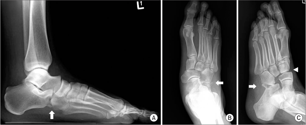
Figure 2 (A, B) Initial anteroposterior and lateral radiograph shows the plantar displacement of the cuboid bone with fracture of the anterior process of the calcaneus (white arrows). (C) Initial oblique radiograph shows navicular-medial cuneiform fracture and dislocation on lateral side (arrowhead), also shows that calcaneocuboid joint is impacted and inferolateral dislocation (white arrow).

Figure 3 (A) The sagittal computed tomography (CT) imaging shows the fracture of calcaneocuboid joint and inferolateral dislocation of cuboid bone. (B) Three-dimensional reconstructed CT scan shows lateral displacement of medial and intermediate cuneiform bone.

Figure 4 (A) Before the anatomical reduction and fixation, we can find calcaneocuboid joint fracture and dislocation in medial column. (B) After definite fixation of medial and lateral column, calcaneocuboid joint is stabilized.

Figure 5 (A) Intraoperative radiograph made after provisional fixation of the medial column and definitive fixation of the lateral column. (B) The radiograph shows anatomical reduction of the length and shape of lateral column.
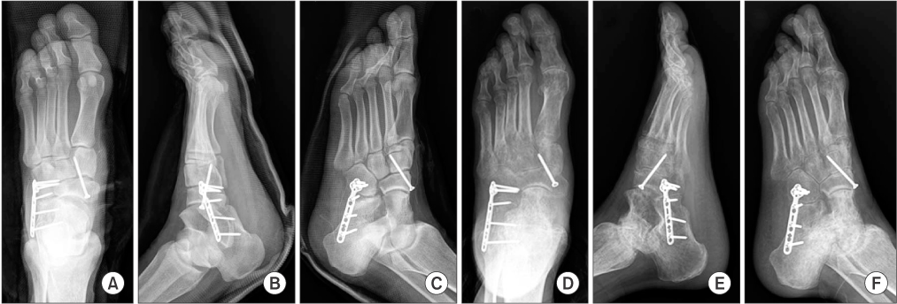
Figure 6 (A~C) Immediate postoperative radiographs show anatomical reduction and stabilization of the midtarsal joint. (D~F) Follow-up radiographs, made at six months, show disuse osteoporosis of the foot.
Reference
-
1. Korean Foot and Ankle Society. The foot and ankle. Seoul: Jin Publishing Co.;2010. p. 26–27.2. The Korean Fracture Society. Principles of fracture management. Seoul: PanMun Education;2013. p. 837–839.3. Richter M, Thermann H, Huefner T, Schmidt U, Goesling T, Krettek C. Chopart joint fracture-dislocation: initial open reduction provides better outcome than closed reduction. Foot Ankle Int. 2004; 25:340–348.
Article4. van Dorp KB, de Vries MR, van der Elst M, Schepers T. Chopart joint injury: a study of outcome and morbidity. J Foot Ankle Surg. 2010; 49:541–545.
Article5. Main BJ, Jowett RL. Injuries of the midtarsal joint. J Bone Joint Surg Br. 1975; 57:89–97.
Article6. Benirschke SK, Meinberg E, Anderson SA, Jones CB, Cole PA. Fractures and dislocations of the midfoot: Lisfranc and Chopart injuries. J Bone Joint Surg Am. 2012; 94:1325–1337.
Article7. Choi JW, Choi JC, Na HY, Shim DJ, Kim YH, Lee SH, et al. Chorpart's dislocation: a case report. J Korean Foot Ankle Soc. 2005; 9:121–124.8. Ip KY, Lui TH. Isolated dorsal midtarsal (Chopart) dislocation: a case report. J Orthop Surg (Hong Kong). 2006; 14:357–359.
Article9. Ly TV, Coetzee JC. Treatment of primarily ligamentous Lisfranc joint injuries: primary arthrodesis compared with open reduction and internal fixation. A prospective, randomized study. J Bone Joint Surg Am. 2006; 88:514–520.
Article
- Full Text Links
-
- Actions
-
Cited
- CITED
-
- Close
- Share
-
- Similar articles
-
- Isolated Plantar Midtarsal Dislocation: A case Report
- Chorpart's Dislocation: A Case Report
- Dislocation of Fifth Carpornetacarpal Joint: Two Cases Report
- Treatment of Traumatic Sternoclavicular Joint Anterior Dislocation with a Sternal Fracture
- Clavicle Midshaft Fracture with Acromioclavicular Joint Dislocation: A Case Report
