J Korean Foot Ankle Soc.
2017 Sep;21(3):79-82. 10.14193/jkfas.2017.21.3.79.
Cause of Metatarsalgia
- Affiliations
-
- 1Department of Orthopedic Surgery, Busan Paik Hospital, Inje University College of Medicine, Busan, Korea.
- 2Department of Orthopedic Surgery, Pusan Medical Center, Busan, Korea. doctordj@paran.com
- KMID: 2389935
- DOI: http://doi.org/10.14193/jkfas.2017.21.3.79
Abstract
- Metatarsalgia is one of the most common causes of patients complaining of pain in their feet. This pain is the plantar forefoot, including the second to fourth metatarsal heads and arises from either mechanical or iatrogenic causes. On the other hand, it is frequently accompanied by a deformity of the toes as well as of the first and fifth rays. The pain has a variety of causes, and sometimes the cause is difficult to distinguish. The variability of possible causative factors necessitates an individualized approach to treatment. To determine these causes, this paper presents an overview of the gait mechanics, plantar pressure, and the classification according to the etiology.
Keyword
MeSH Terms
Figure
-
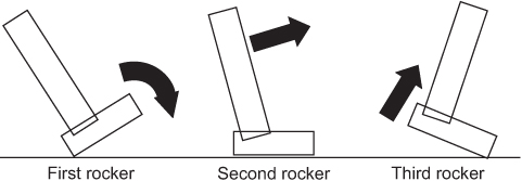
Figure 1 Event of three rocker phases of walking. The heel acts as the first rocker, beginning with initial heel strike. In the second rocker, the entire foot normally remains in contact with the ground. During the third rocker only the forefoot is in contact with the ground.
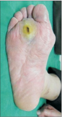
Figure 2 An elevated first metatarsus pushes the whole load of the second rocker onto the second metatarsus, resulting in isolated keratosis underneath the second metatarsal head.
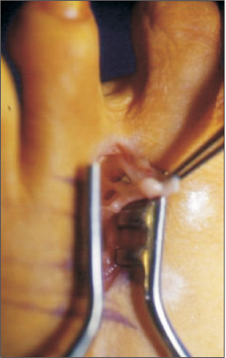
Figure 3 This photo shows Morton's neuroma between 3rd~4th metatarsal heads.
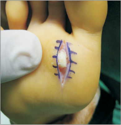
Figure 4 This photo shows benign tumor underneath the second metatarsal head.
Reference
-
1. Espinosa N, Maceira E, Myerson MS. Current concept review: metatarsalgia. Foot Ankle Int. 2008; 29:871–879.
Article2. Perry J, Schoneberger B. Gait analysis normal and pathological function. Thorofare: Slack;1992.3. Göpfert B, Valderrabano V, Hintermann B, Wirz D. Measurement of the isometric dorsiflexion and plantar flexion force in the ankle joint. Biomed Tech (Berl). 2005; 50:282–286.
Article4. Trnka HJ, Gebhard C, Mühlbauer M, Ivanic G, Ritschl P. The Weil osteotomy for treatment of dislocated lesser metatarsophalangeal joints: good outcome in 21 patients with 42 osteotomies. Acta Orthop Scand. 2002; 73:190–194.
Article5. Aronow MS, Diaz-Doran V, Sullivan RJ, Adams DJ. The effect of triceps surae contracture force on plantar foot pressure distribution. Foot Ankle Int. 2006; 27:43–52.
Article6. Kirtley C. Clinical gait analysis: theory and practice. Edinburgh: Elsevier;2006.7. Besse JL. Metatarsalgia. Orthop Traumatol Surg Res. 2017; 103(1S):S29–S39.
Article8. Cavanagh PR, Rodgers MM, Iiboshi A. Pressure distribution under symptom-free feet during barefoot standing. Foot Ankle. 1987; 7:262–276.
Article9. Espinosa N, Brodsky JW, Maceira E. Metatarsalgia. J Am Acad Orthop Surg. 2010; 18:474–485.
Article10. Maestro M, Besse JL, Ragusa M, Berthonnaud E. Forefoot morphotype study and planning method for forefoot osteotomy. Foot Ankle Clin. 2003; 8:695–710.
Article11. Thompson IM, Bohay DR, Anderson JG. Fusion rate of first tarsometatarsal arthrodesis in the modified Lapidus procedure and flatfoot reconstruction. Foot Ankle Int. 2005; 26:698–703.
Article12. Crosbie J, Burns J, Ouvrier RA. Pressure characteristics in painful pes cavus feet resulting from Charcot-Marie-Tooth disease. Gait Posture. 2008; 28:545–551.
Article13. Fuhrmann RA. Lesser toe deformities. Ther Umsch. 2004; 61:417–420.14. Hansen ST. Functional reconstruction of the foot and ankle. Philadelphia: Lippincott Williams & Wilkins;2000. p. 145–186.15. Jung HG, Zaret DI, Parks BG, Schon LC. Effect of first metatarsal shortening and dorsiflexion osteotomies on forefoot plantar pressure in a cadaver model. Foot Ankle Int. 2005; 26:748–753.
Article16. Ledoux WR, Shofer JB, Ahroni JH, Smith DG, Sangeorzan BJ, Boyko EJ. Biomechanical differences among pes cavus, neutrally aligned, and pes planus feet in subjects with diabetes. Foot Ankle Int. 2003; 24:845–850.
Article17. Yu GV, Judge MS, Hudson JR, Seidelmann FE. Predislocation syndrome Progressive subluxation/dislocation of the lesser metatarsophalangeal joint. J Am Podiatr Med Assoc. 2002; 92:182–199.18. Myerson MS, Jung HG. The role of toe flexor-to-extensor transfer in correcting metatarsophalangeal joint instability of the second toe. Foot Ankle Int. 2005; 26:675–679.
Article19. Coughlin MJ. Common causes of pain in the forefoot in adults. J Bone Joint Surg Br. 2000; 82:781–790.
Article20. Fortin PT, Myerson MS. Second metatarsophalangeal joint instability. Foot Ankle Int. 1995; 16:306–313.
Article21. Vora AM, Myerson MS. First metatarsal osteotomy nonunion and malunion. Foot Ankle Clin. 2005; 10:35–54.
Article22. Acevedo JI. Fixation of metatarsal osteotomies in the treatment of hallux valgus. Foot Ankle Clin. 2000; 5:451–468.23. Espinosa N, Myerson MS, Fernandez De Retana P, Maceira E. A new approach for the treatment of metatarsalgia: the triple Weil osteotomy. Tech Foot Ankle Surg. 2007; 6:254–263.24. Maceira E, Monteagudo M. Transfer metatarsalgia post hallux valgus surgery. Foot Ankle Clin. 2014; 19:285–307.
Article
- Full Text Links
-
- Actions
-
Cited
- CITED
-
- Close
- Share
-
- Similar articles
-
- Treatment of Metatarsalgia after a Surgery
- Lesser Metatarsal Osteotomies for Metatarsalgia
- A problem-based approach in musculoskeletal ultrasonography: central metatarsalgia
- Modified Scarf Osteotomy for Hallux Valgus with Lesser Metatarsalgia
- A Clinical Study of Chevron Osteotomy in Bunion - Hallux Valgus
