Diseases treated in oculoplasty
- Affiliations
-
- 1Department of Ophthalmology, St. Vincent's Hospital, The Catholic University of Korea College of Medicine, Suwon, Korea. laty@catholic.ac.kr
- KMID: 2389790
- DOI: http://doi.org/10.5124/jkma.2017.60.9.719
Abstract
- Most people think that ophthalmology only treat diseases of the eyeball, but there are also many diseases of the accessory organs around the eyeball. Oculoplasty is a field of ophthalmology that deals with diseases of the eyelids, lacrimal system, and orbit. These accessory organs play important roles in protecting the eyes and supporting their function. Sometimes, diseases of these organs cause cosmetic problems, as well as functional problems. In the past, this field was considered rather indifferently and was not well recognized, so the treatment of these diseases was not specialized. However, concomitantly with recent improvements in quality of life and the increased desire of patients for these diseases to be treated, the importance of this field is increasing and many oculoplastic specialists are being trained. In the oculoplastic field, wide range of diseases are treated. In this report, the author provides a brief overview of the most important and common oculoplastic diseases.
MeSH Terms
Figure
-

Figure 1 Epiblepharon.
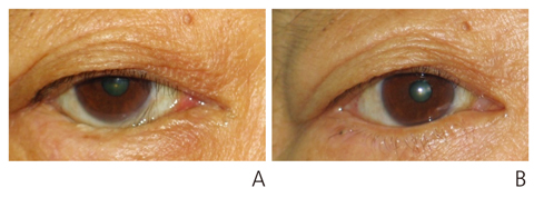
Figure 2 Entropion of lower lid. (A) Preoperative and (B) postoperative photograph.
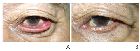
Figure 3 Ectropion of lower lid. (A) Preoperative and (B) postoperative photograph.

Figure 4 Congenital ptosis. (A) Preoperative and (B) postoperative photograph of frontalis sling.
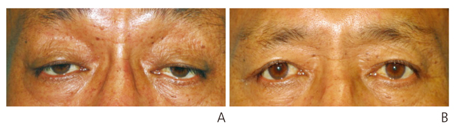
Figure 5 Acquired ptosis. (A) Preoperative and (B) postoperative photograph of levator resection.
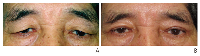
Figure 6 Dermatochalasis. (A) Preoperative and (B) postoperative photograph.
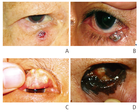
Figure 7 Malignant tumors of lid. (A) Basal cell carcinoma, (B) squamous cell carcinoma, (C) sebaceous cell carcinoma, and (D) malignant melanoma.
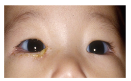
Figure 8 Congenital nasolacrimal duct obstruction.
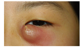
Figure 9 Acute dacryocystitis.
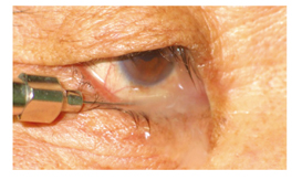
Figure 10 Chronic dacryocystitis.

Figure 11 Acute canaliculitis. (A) Pouting punctum and (B) sulfur granule.
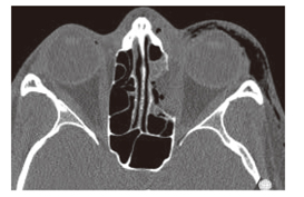
Figure 12 Blow out fracture (medial wall, left).
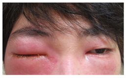
Figure 13 Preseptal cellulitis.
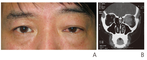
Figure 14 Pseudotumor of orbit. (A) Proptosis and conjunctival injection of left eye. (B) Diffuse infiltration of inflammatory mass in left orbit.
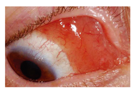
Figure 15 Conjunctival lymphoma.
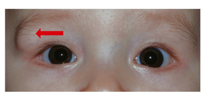
Figure 16 Dermoid cyst (right).

Figure 17 (A) Intraoperative photograph of orbital cavernous hemangioma removal. (B) Completely removed cavernous hemangioma.
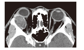
Figure 18 Pleomorphic adenoma of lacrimal gland in right orbit.
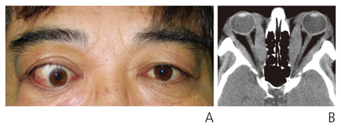
Figure 19 Thyroid associated ophthalmopathy. (A) Proptosis and hypotropia of right eye. (B) Orbit computed tomography shows severe hypertrophy of medial rectus muscle.
Cited by 1 articles
-
Introduction of ophthalmic plastic and reconstructive surgery
Suk-Woo Yang
J Korean Med Assoc. 2017;60(9):717-718. doi: 10.5124/jkma.2017.60.9.717.
Reference
-
1. Kim HM, Lee TS. Clinical observation and their surgical results of 127 cases. J Korean Ophthalmol Soc. 1985; 26:441–448.2. Jung W, Lee OS. A clinical study on surgical results of frontalis sling in relation to the operative age. J Korean Ophthalmol Soc. 1999; 40:1368–1374.3. Kim KH, Chung WS. Classification and therapeutic effect of benign and malignant eyelid tumors. J Korean Ophthalmol Soc. 1997; 38:703–709.4. Lee HK, Hu CH, Kkkang SJ, Lee SY. Clinical analysis of lid tumors. J Korean Ophthalmol Soc. 1997; 38:1892–1898.5. Lee TS, Lee JJ. Analysis of classification and incidence of eyelid and orbital tumors. J Korean Ophthalmol Soc. 1997; 38:1700–1705.6. Korean Society of Oculoplastic and Reconstructive Surgery. Ophthalmic plastic and reconstructive surgery. 3rd ed. Goyang: Naewaehaksool;2015.7. Horie C, Suzuki Y, Kiyosawa M, Mochizuki M, Wakakura M, Oda K, Ishiwata K, Ishii K. Decreased dopamine D receptor binding in essential blepharospasm. Acta Neurol Scand. 2009; 119:49–54.8. Micheli F, Scorticati MC, Folgar S, Gatto E. Development of Parkinson's disease in patients with blepharospasm. Mov Disord. 2004; 19:1069–1072.
Article9. Ross AH, Elston JS, Marion MH, Malhotra R. Review and update of involuntary facial movement disorders presenting in the ophthalmological setting. Surv Ophthalmol. 2011; 56:54–67.
Article10. Lee SY, Chung HS, Kim HB, Namgung R, Han DG. The incidence of congenital nasolacrimal duct obstruction in Korean neonates. J Korean Ophthalmol Soc. 1989; 30:5–8.11. Oh HS, Ahn Y. The incidence and medical treatment of congenital nasolacrimal duct obstruction in Korean infants. J Korean Ophthalmol Soc. 1995; 36:1007–1013.12. Donahue SP, Schwartz G. Preseptal and orbital cellulitis in childhood: a changing microbiologic spectrum. Ophthalmolog. 1998; 105:1902–1905.13. Schwartz GR, Wright SW. Changing bacteriology of periorbital cellulitis. Ann Emerg Med. 1996; 28:617–620.
Article14. American Academy of Ophthalmology. Orbit, eyelid, lacrimal system: Basic and Clinical Science Course 2007-2008. San Francisco: American Academy of Ophthalmology;2007.15. Bahn RS. Graves' ophthalmopathy. N Engl J Med. 2010; 362:726–738.
Article16. Garrity JA, Bahn RS. Pathogenesis of Graves' ophthalmopathy: implications for prediction, prevention, and treatment. Am J Ophthalmol. 2006; 142:147–153.
Article17. Rhee KC, Lee TS. The clinical study on Graves' ophthalmopathy. J Korean Ophthalmol Soc. 1999; 40:2923–2927.18. Woo KI, Kim YD, Lee SY. Korean Society of Ophthalmic Plastic and Reconstructive Surgery. The clinical characteristics of thyroid orbitopathy in thyroid dysfunction patients in Korea. J Korean Ophthalmol Soc. 2008; 49:1387–1396.
Article
- Full Text Links
-
- Actions
-
Cited
- CITED
-
- Close
- Share
-
- Similar articles
-
- Guardians' Perceptions of Pediatric Oculoplasty: Comparative Analysis in 2007 and 2017
- Isolated Unilateral Tessier Cleft 10 with Anterior Staphyloma
- Present and future of oculoplasty
- Rare and Atypical Presentation of Glomus Tumor in the Eyelid
- The Morphological and Functional Characteristics of Eyelid in Korean Children
