Ann Rehabil Med.
2017 Aug;41(4):589-594. 10.5535/arm.2017.41.4.589.
Anatomical Localization of Motor Points of the Abductor Hallucis Muscle: A Cadaveric Study
- Affiliations
-
- 1Department of Rehabilitation Medicine, Seoul St. Mary's Hospital, College of Medicine, The Catholic University of Korea, Seoul, Korea. drpjh@catholic.ac.kr
- 2Rhin Hospital, Yongin, Korea.
- 3Department of Rehabilitation Medicine, Eulji General Hospital, Seoul, Korea.
- KMID: 2389404
- DOI: http://doi.org/10.5535/arm.2017.41.4.589
Abstract
OBJECTIVE
To identify the anatomical motor points of the abductor hallucis muscle in cadavers.
METHODS
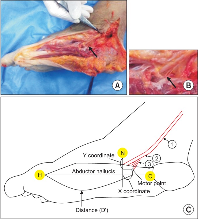
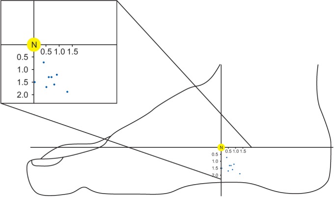
Motor nerve branches to the abductor hallucis muscles were examined in eight Korean cadaver feet. The motor point was defined as the site where the intramuscular nerve penetrates the muscle belly. The reference line connects the metatarsal base of the hallux (H) to the medial tubercle of the calcaneus (C). The x coordinate was the horizontal distance from the motor point to the point where the perpendicular line from the navicular tuberosity crossed the reference line. The y coordinate was the perpendicular distance from the motor point to the navicular tuberosity.
RESULTS
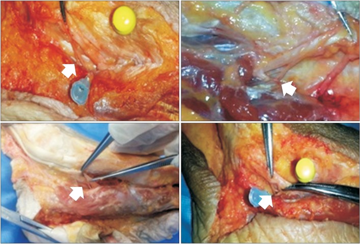
Most of the medial plantar nerves to the abductor hallucis muscles divide into multiple branches before entering the muscles. One, two, and three motor branches were observed in 37.5%, 37.5%, and 25% of the feet, respectively. The ratios of the main motor point from the H with respect to the H-C line were: main motor point, 68.79%±5.69%; second motor point, 73.45%±3.25%. The mean x coordinate value from the main motor point was 0.65±0.49 cm. The mean value of the y coordinate was 1.43±0.35 cm. All of the motor points of the abductor hallucis were consistently found inferior and posterior to the navicular tuberosity.
CONCLUSION
This study identified accurate locations of anatomical motor points of the abductor hallucis muscle by means of cadaveric dissection, which can be helpful for electrophysiological studies in order to correctly diagnose the various neuropathies associated with tibial nerve components.
Keyword
Figure
Reference
-
1. Dumitru D, Amato AA, Zwarts MJ. Electrodiagnostic medicine. Philadelphia: Hanley & Belfus;2002.2. Liveson JA, Ma DM. Laboratory reference for clinical neurophysiology. New York: Oxford University Press;1992. p. 204–205.3. Lee HJ, DeLisa JA, Lee HJ. Manual of nerve conduction study and surface anatomy for needle electromyography. Philadelphia: Lippincott Williams & Wilkins;2005. p. 80–83.4. Pease WS, Lew HL, Johnson EW. Johnson's practical electromyography. Philadelphia: Lippincott Williams & Wilkins;2007. p. 242.5. Del Toro DR, Park TA. Abductor hallucis false motor points: electrophysiologic mapping and cadaveric dissection. Muscle Nerve. 1996; 19:1138–1143. PMID: 8761270.
Article6. Bromberg MB, Spiegelberg T. The influence of active electrode placement on CMAP amplitude. Electroencephalogr Clin Neurophysiol. 1997; 105:385–389. PMID: 9363004.
Article7. Downey JA, Myers SJ, Gonzalez EG, Lieberman JS. The physiological basis of rehabilitation medicine. 2nd ed. Boston: Butterworth-Heinemann;1994. p. 576.8. van Dijk JG, van Benten I, Kramer CG, Stegeman DF. CMAP amplitude cartography of muscles innervated by the median, ulnar, peroneal, and tibial nerves. Muscle Nerve. 1999; 22:378–389. PMID: 10086899.
Article9. Rodriguez-Carreno I, Malanda-Triqueros A, Gila-Useros L. Motor unit action potential duration: measurement and significance. In : Ajeena IM, editor. Advances in clinical neurophysiology. Rijeka: InTech;2012. p. 133–160.10. Brashear A, Kincaid JC. The influence of the reference electrode on CMAP configuration: leg nerve observations and an alternative reference site. Muscle Nerve. 1996; 19:63–67. PMID: 8538671.
Article11. Park TA, Jurell KC, Del Toro DR. Generator sources for the early and late ulnar hypothenar premotor potentials: short segment electrophysiologic studies and cadaveric dissection. Arch Phys Med Rehabil. 1996; 77:467–472. PMID: 8629923.
Article12. Bilbao A, Wilcox MS. The influence of the reference electrode on CMAP configuration: leg nerve observations and an alternative reference site. Muscle Nerve. 1996; 19:1055–1056. PMID: 8756176.
Article13. Monteiro A, Cricenti SV, Manzano GM, Nobrega JA. Anatomical observations and false motor points in intrinsic foot muscles. Muscle Nerve. 2002; 26:557. PMID: 12362425.
Article14. del Sol M, Olave E, Gabrielli C, Mandiola E, Prates JC. Innervation of the abductor digiti minimi muscle of the human foot: anatomical basis of the entrapment of the abductor digiti minimi nerve. Surg Radiol Anat. 2002; 24:18–22. PMID: 12197005.
Article15. Wong YS. Influence of the abductor hallucis muscle on the medial arch of the foot: a kinematic and anatomical cadaver study. Foot Ankle Int. 2007; 28:617–620. PMID: 17559771.
Article16. Fiolkowski P, Brunt D, Bishop M, Woo R, Horodyski M. Intrinsic pedal musculature support of the medial longitudinal arch: an electromyography study. J Foot Ankle Surg. 2003; 42:327–333. PMID: 14688773.
Article17. Dumitru D, DeLisa JA. AAEM Minimonograph #10: volume conduction. Muscle Nerve. 1991; 14:605–624. PMID: 1922167.
Article18. Aquilonius SM, Askmark H, Gillberg PG, Nandedkar S, Olsson Y, Stalberg E. Topographical localization of motor endplates in cryosections of whole human muscles. Muscle Nerve. 1984; 7:287–293. PMID: 6203034.
Article
- Full Text Links
- Actions
-
Cited
- CITED
-
- Close
- Share
- Similar articles
-
- Influence of Longitudinal Arch of Foot on the Strength and Muscle Activity of the Abductor Hallucis in Subjects with and without Navicular Drop Sign
- Availability of Neurophysiological Index in Amyotrophic Lateral Sclerosis: Application of Neurophysiological Index to Median Nerve /Abductor Digiti Minimi and Posterior Tibial Nerve/Abductor Hallucis
- The Checkrein Deformity of Extensor Hallucis Longus Tendon and Extensor Retinaculum Syndrome with Deep Peroneal Nerve Entrapment after Triplane Fracture: A Case Report
- The Significance of Motor Evoked Potentials as a Prognostic Factor in the Early Stage of Stroke Patients
- Interictal Motor Cortex Excitability in Migraine and Chronic Tension Type Headache using Transcranial Magnetic Stimulation


