Ann Dermatol.
2017 Aug;29(4):499-500. 10.5021/ad.2017.29.4.499.
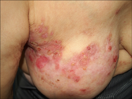
A Case of Wolf's Isotopic Response Presenting as Bullous Pemphigoid
- Affiliations
-
- 1Department of Dermatology, Korea University Ansan Hospital, College of Medicine, Korea University, Ansan, Korea. dermhj@naver.com
- KMID: 2388955
- DOI: http://doi.org/10.5021/ad.2017.29.4.499
Abstract
- No abstract available.
MeSH Terms
Figure
Reference
-
1. Ruocco V, Ruocco E, Brunetti G, Russo T, Gambardella A, Wolf R. Wolf's post-herpetic isotopic response: Infections, tumors, and immune disorders arising on the site of healed herpetic infection. Clin Dermatol. 2014; 32:561–568.
Article2. Noh TW, Park SH, Kang YS, Lee UH, Park HS, Jang SJ. Morphea developing at the site of healed herpes zoster. Ann Dermatol. 2011; 23:242–245.
Article3. Gurel MS, Savas S, Bilgin F, Erdil D, Leblebici C, Sarikaya E. Zosteriform pemphigoid after zoster: Wolf's isotopic response. Int Wound J. 2016; 13:141–142.
Article4. Fichel F, Barbe C, Joly P, Bedane C, Vabres P, Truchetet F, et al. Clinical and immunologic factors associated with bullous pemphigoid relapse during the first year of treatment: a multicenter, prospective study. JAMA Dermatol. 2014; 150:25–33.
Article5. Kamiya K, Aoyama Y, Suzuki T, Niwa H, Horio A, Nishio E, et al. Possible enhancement of BP180 autoantibody production by herpes zoster. J Dermatol. 2016; 43:197–199.
Article
- Full Text Links
- Actions
-
Cited
- CITED
-
- Close
- Share
- Similar articles
-
- Coexistence of Bullous Pemphigoid and Psoriasis: A Case Report and Review of the Literature
- A Case of Bullous Pemphigoid Associated with Prostate Adenocarcinoma
- A Case of Dyshidrosiform Pemphigoid
- A Case of Benign Fibrous Histiocytoma on Herpes Zoster Scar: Wolf's Isotopic Response
- Multiple Epidermal Cysts after Herpes Zoster: Wolf's Isotopic Response


