Prog Med Phys.
2017 Jun;28(2):45-48. 10.14316/pmp.2017.28.2.45.
Assessment of Radiation Dose from Radioactive Wedge Filters during High-Energy X-Ray Therapy
- Affiliations
-
- 1Department of Radiation Oncology, Asan Medical Center, Seoul, Korea.
- 2Department of Neurosurgery, Ulsan University Hospital, Ulsan, Korea. michael@uuh.ulsan.kr
- 3Department of Radiological Science, Kangwon National University, Samcheok, Korea.
- KMID: 2388207
- DOI: http://doi.org/10.14316/pmp.2017.28.2.45
Abstract
- This paper evaluated the amount of radiation generated by wedge filters during radiation therapy using a high-energy linear accelerator, and the dose to the worker during wedge replacement. After 10-MV photon beam was irradiated with wedge filter, the wedge was removed from the linear accelerator, and the dose rate and energy spectrum were measured. The initial measurement was approximately 1 uSv/h, and the radiation level was reduced to 0.3 uSv/h after 6 min. The effective half-life derived from the dose rate measurement was approximately 3.5 min, and the influence of AI-28 was about 53%. From the energy spectrum measurements, a peak of 1,799 keV was measured for AI-28, while the peak for Co-58 was not measured in the control room. The peaks for Au-106 and Cd-105 were found only measurement was done without wedge removement from the linear accelerator. The additional doses received by the radiation worker during wedge replacement were estimated to be 0.08−0.4 mSv per year.
Keyword
MeSH Terms
Figure
-
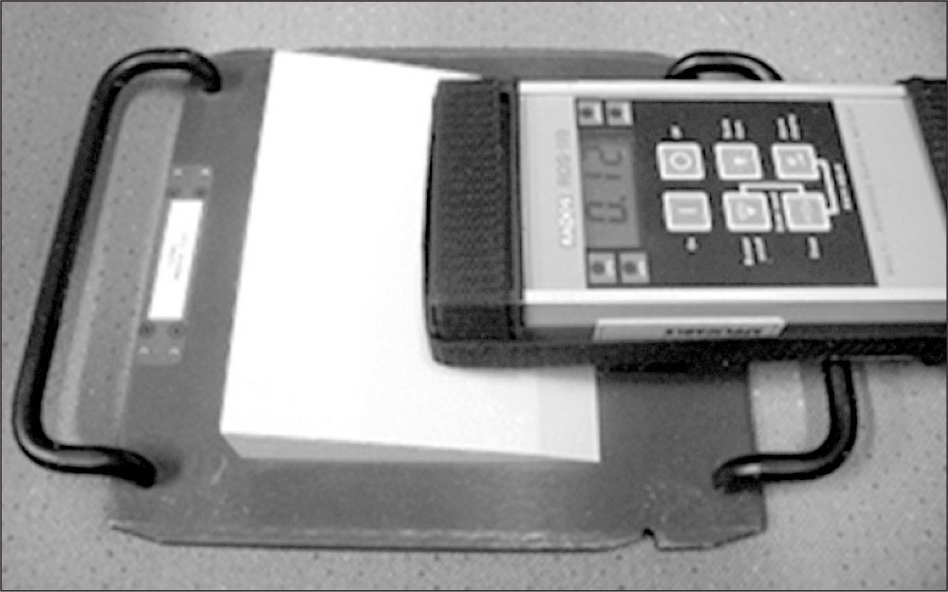
Fig. 1. Measurement of dose rate using GM detector and activated wedge filter.
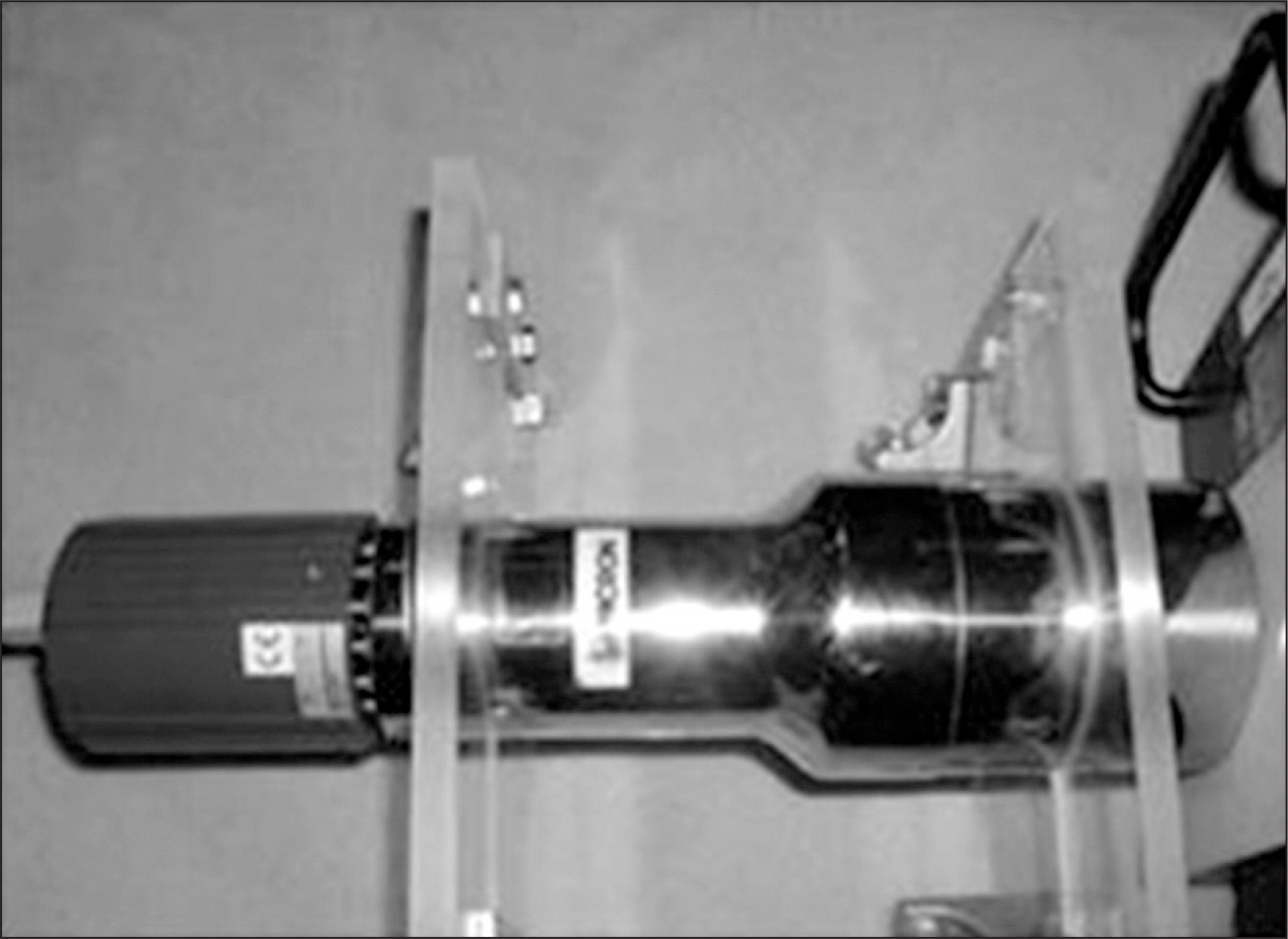
Fig. 2. Measurement of energy spectrum using NaI scintillator detector.
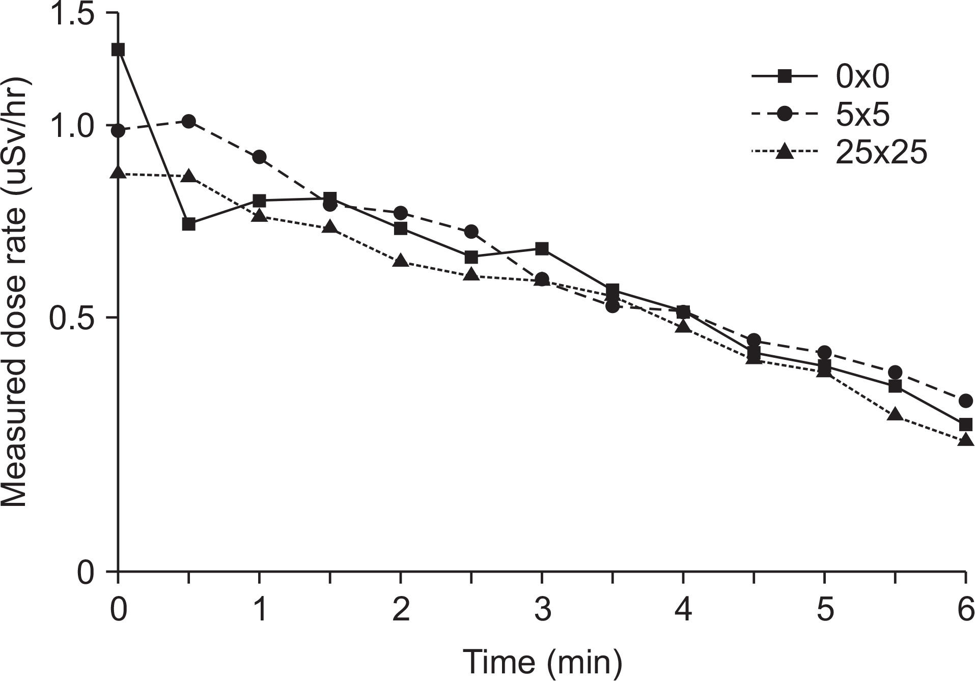
Fig. 3. Measured dose rate of activated wedge after 5-Gy irradiation at various field sizes.
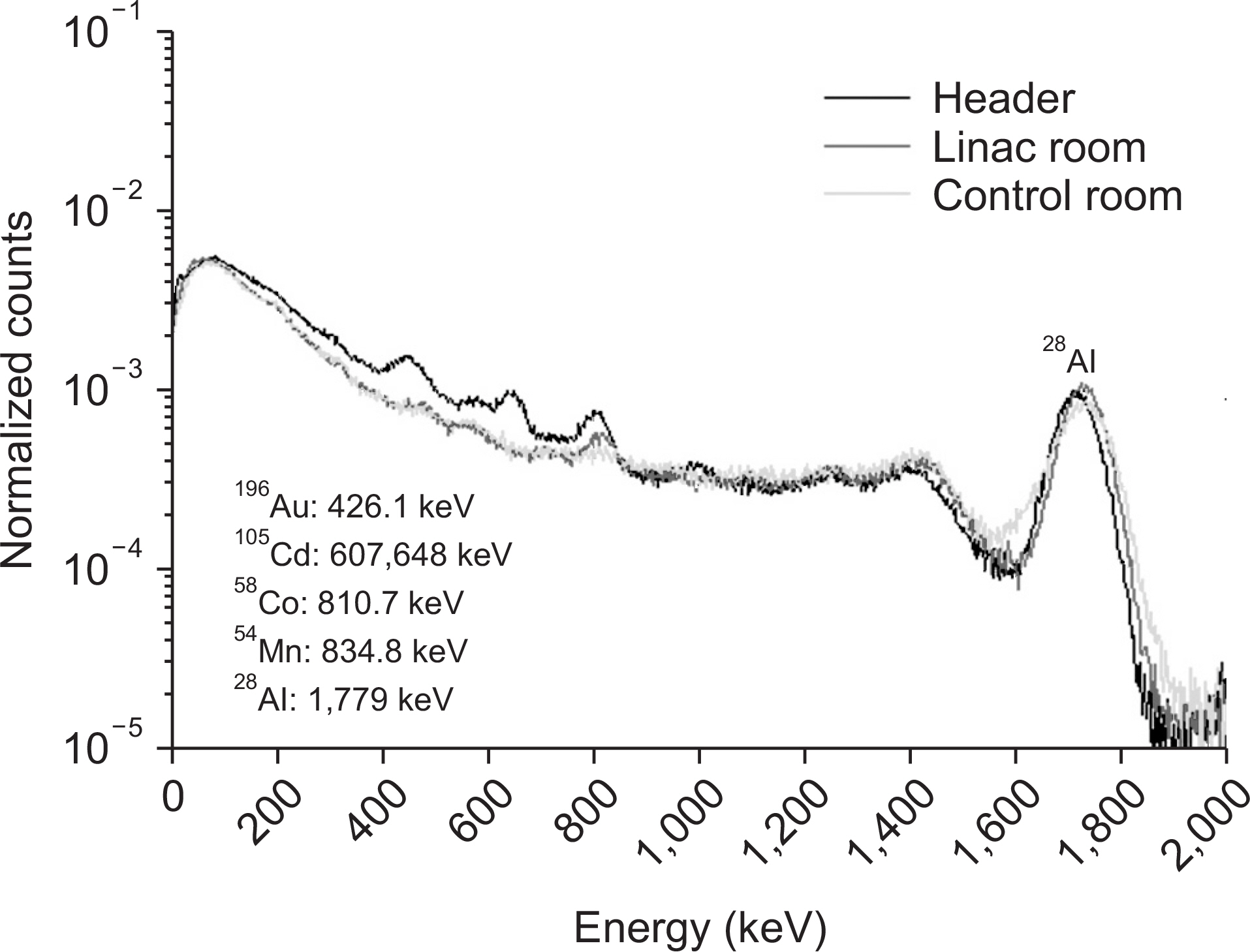
Fig. 4. Measured energy spectrum from activated wedge after 5-Gy irradiation.
Reference
-
1. NCRP Report 79. Neutron contamination from medical electron accelerator. 1984.2. AAPM TG Report 27. Neutron measurements around high energy X-ray radiotherapy machines. 1986.3. Chilbani O, Ma C. Photoneutron dose calculations for high energy photon beams from Siemens and Varian LINACS. Med Phys 1990–2000. 2003; 30.4. Gudowska I, Brahme A, Andreo P, et al. Calcluation of absorbed dose and biological effectiveness from photoneutron reactions in bremsstrahlungbeam of end point 50 MeV. Phys in Med Biol. 1999; 44:2099.5. Roig M, Panettieri V, Ginjaume M, et al. Photonuclear isotope characterization of a Siemens KDS 18 MeV LINAC head. Phys in Med Biol. 2004; 49:N242–6.6. Borchardt IM, Patterson JR, Beddoe AH. An investigation of photonuclear reactions in Cerrobend eutectic material with an 18 MV LINAC. Phys in Med Biol. 1991; 36:649–53.
Article7. Petrovic N, Krestic-Vesovic J, Stojanovic D, et al. Contribution of activation products to occupational exposure following treatment using high energy photons in radiotherapy. Rad Prot Dos. 2011; 143:109–12.8. Fischer HW, Tabot BE, Poppe B. Activation processes in a medical linear accelerator and spatial distribution of activation products. Phys Med Biol. 2006; 51:N461.
Article9. Fischer HW, Tabot B, Poppe B. Comparison of activation products and induced dose rates in diferent high-energy medical linear accelerators. Health Physics. 2008; 94:272–8.10. Israngkul-Na-Ayuthaya I, Suriyapee S, Pengvanich P. Evaluation of equivalent dose from neutrons and activation products from a 15 MV x-ray LINAC. J of Rad Research. 2015; 56:919–26.
- Full Text Links
-
- Actions
-
Cited
- CITED
-
- Close
- Share
-
- Similar articles
-
- A Study on Dose Distribution using Virtual Wedge in Breast Cancer
- Dosimetric Characteristics of Dynamic Wedge Techinique
- Film Dosimetry for Intensity Modulated Radiation Therapy: Dosimetric Evaluation
- Simple design of wedge filters for Co teletherapy
- The Enhancement of Skin Sparing by Tray Materials for High Energy Photon Beam
