J Dent Rehabil Appl Sci.
2017 Jun;33(2):119-126. 10.14368/jdras.2017.33.2.119.
The effect of Light Emitting Diode electric toothbrush on gingivitis: a randomized controlled trial
- Affiliations
-
- 1Department of Periodontology, College of Dentistry, Dankook University, Cheonan, Republic of Korea. periolee85@gmail.com
- KMID: 2388018
- DOI: http://doi.org/10.14368/jdras.2017.33.2.119
Abstract
- PURPOSE
The aim of the present study was to compare clinical antiplaque and antigingivitis effect between Light Emitting Diode (LED) electronic toothbrush and electronic toothbrush without LED for gingivitis and mild periodontitis patients.
MATERIALS AND METHODS
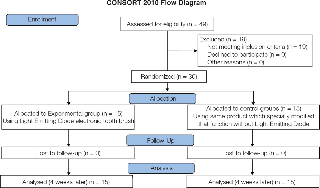
30 patients included in this study. 15 patients in experimental group used LED electronic tooth brush which has red and white LED within its head, and other 15 patients in control group used same product which specially modified that function without LED. Clinical parameters (Löe-Silness gingival index (GI), Quigley-Hein plaque index (PI)) were measured at the baseline, 2 weeks and 4 weeks later. Wilcoxon signed rank test and Mann-Whitney test were used for statistical analysis.
RESULTS
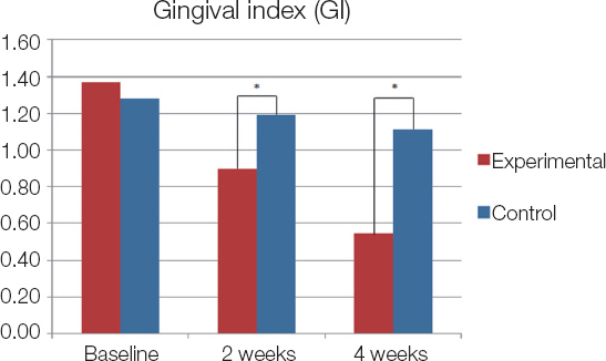
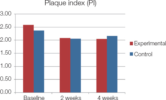
Compare of GI change between experimental and control group with time, both groups showed that reduced GI, but lower GI values detected at 2 weeks and 4 weeks later in experimental group than control group. And lower PI values detected at 4 weeks later in experimental group than control group, but not statistically significant.
CONCLUSION
Based on these results and within the limits of this study, the electronic toothbrush with LED could reducing gingivitis in a short period and infer that decreasing plaque accumulation in a long period.
Keyword
MeSH Terms
Figure
Reference
-
References
1. Becker W, Becker BE, Berg LE. Periodontal treatment without maintenance. A retrospective study in 44 patients. J Periodontol. 1984; 55:505–9. DOI: 10.1902/jop.1984.55.9.505. PMID: 6592322.2. Lindhe J, Westfelt E, Nyman S, Socransky SS, Haffajee AD. Long-term effect of surgical/nonsurgical treatment of periodontal disease. J Clin Periodontol. 1984; 11:448–58. DOI: 10.1111/j.1600-051X.1984.tb00902.x. PMID: 6378986.3. Silverstone LM, Featherstone MJ. A scanning electron microscope study of the end rounding of bristles in eight toothbrush types. Quintessence Int. 1988; 19:87–107.4. Hawkins D, Abrahamse H. Phototherapy-a treatment modality for wound healing and pain relief. African J Biomed Res. 2007; 10:99–109.5. Wong-Riley MT, Bai X, Buchmann E, Whelan HT. Light-emitting diode treatment reverses the effect of TTX on cytochrome oxidase in neurons. Neuroreport. 2001; 12:3033–7. DOI: 10.1097/00001756-200110080-00011. PMID: 11568632.6. Whelan HT, Connelly JF, Hodgson BD, Barbeau L, Post AC, Bullard G, Buchmann EV, Kane M, Whelan NT, Warwick A, Margolis D. NASA lightemitting diodes for the prevention of oral mucositis in pediatric bone marrow transplant patients. J Clin Laser Med Surg. 2002; 20:319–24. DOI: 10.1089/104454702320901107. PMID: 12513918.7. Whelan HT, Houle JM, Donohoe DL, Bajic DM, Schmidt MH, Reichert KW, Weyenberg GT, Larson DL, Meyer GA, Caviness JA. Medical applications of space light-emitting diode technology - space station and beyond. AIP Conference Proceedings. 1999; 458:3–16. DOI: 10.1063/1.57600.8. Whelan HT, Smits RL Jr, Buchman EV, Whelan NT, Turner SG, Margolis DA, Cevenini V, Stinson H, Ignatius R, Martin T, Cwiklinski J, Philippi AF, Graf WR, Hodgson B, Gould L, Kane M, Chen G, Caviness J. Effect of NASA light-emitting diode irradiation on wound healing. J Clin Laser Med Surg. 2001; 19:305–14. DOI: 10.1089/104454701753342758. PMID: 11776448.9. Whelan HT, Buchmann EV, Whelan NT, Turner SG, Cevenini V, Stinson H, Ignatius R, Martin T, Cwiklinski J, Meyer GA, Hodgson B, Gould L, Kane M, Chen G, Caviness J. NASA light emitting diode medical applications from deep space to deep sea. AIP Conference Proceedings. 2001; 552:35–45. DOI: 10.1063/1.1357902.10. Löe H, Theilade E, Jensen SB. Experimental gingivitis in man. J Periodontol. 1965; 36:177–87. DOI: 10.1902/jop.1965.36.3.177.11. Lee SY, You CE, Park MY. Blue and red light combination LED phototherapy for acne vulgaris in patients with skin phototype IV. Lasers Surg Med. 2007; 39:180–8. DOI: 10.1002/lsm.20412. PMID: 17111415.12. Lang-Bicudo L, Eduardo Fde P, Eduardo Cde P, Zezell DM. LED phototherapy to prevent mucositis:a case report. Photomed Laser Surg. 2008; 26:609–13. DOI: 10.1089/pho.2007.2228. PMID: 19025412.13. Whelan HT, Buchmann EV, Dhokalia A, Kane MP, Whelan NT, Wong-Riley MT, Eells JT, Gould LJ, Hammamieh R, Das R, Jett M. Effect of NASA light-emitting diode irradiation on molecular changes for wound healing in diabetic mice. J Clin Laser Med Surg. 2003; 21:67–74. DOI: 10.1089/104454703765035484. PMID: 12737646.14. de Sousa AP, Santos JN, Dos Reis JA Jr, Ramos TA, de Souza J, Cangussú MC, Pinheiro AL. Effect of LED phototherapy of three distinct wavelengths on fibroblasts on wound healing:a histological study in a rodent model. Photomed Laser Surg. 2010; 28:547–52. DOI: 10.1089/pho.2009.2605. PMID: 20001321.15. Baez F, Reilly LR. The use of light-emitting diode therapy in the treatment of photoaged skin. J Cosmet Dermatol. 2007; 6:189–94. DOI: 10.1111/j.1473-2165.2007.00329.x. PMID: 17760698.16. Barolet D. Light-emitting diodes (LEDs) in dermatology. Semin Cutan Med Surg. 2008; 27:227–38. DOI: 10.1016/j.sder.2008.08.003. PMID: 19150294.17. Eells JT, DeSmet KD, Kirk DK, Wong-Riley M, Whelan HT, Ver Hoeve J, Nork TM, Stone J, Valter K. Photobiomodulation for the treatment of retinal injury and retinal degenerative diseases. Proceedings of Light-Activated Tissue Regeneration and Therapy Conference. 2008; 39–51. DOI: 10.1007/978-0-387-71809-5_5.18. Erdle BJ, Brouxhon S, Kaplan M, Vanbuskirk J, Pentland AP. Effects of continuous-wave (670-nm) red light on wound healing. Dermatol Surg. 2008; 34:320–5. DOI: 10.1111/j.1524-4725.2007.34065.x. PMID: 18177400 .
- Full Text Links
- Actions
-
Cited
- CITED
-
- Close
- Share
- Similar articles
-
- Effects of an electric toothbrush combined with 3-color light-emitting diodes on antiplaque and bleeding control: a randomized controlled study
- The shear bond strength and adhesive failure pattern in bracket bonding with different light-curing methods
- A comparative study of electric and manual toothbrushes on oral hygiene status in fixed orthodontic patients
- Topical Photodynamic Therapy for Treatment of Actinic Keratosis Using Light-Emitting Diode (LED) Device
- Transfer printing based manufacturing process for flexible micro-light emitting diode phototherapy devices


