J Korean Assoc Oral Maxillofac Surg.
2017 Jun;43(3):191-196. 10.5125/jkaoms.2017.43.3.191.
Squamous cell carcinoma of the buccal mucosa involving the masticator space: a case report
- Affiliations
-
- 1Department of Oral and Maxillofacial Surgery, School of Dentistry, Seoul National University, Seoul, Korea. myoungh@snu.ac.kr
- 2Dental Research Institute, Seoul National University, Seoul, Korea.
- KMID: 2386366
- DOI: http://doi.org/10.5125/jkaoms.2017.43.3.191
Abstract
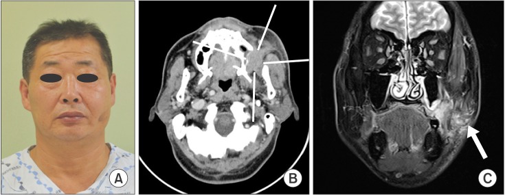
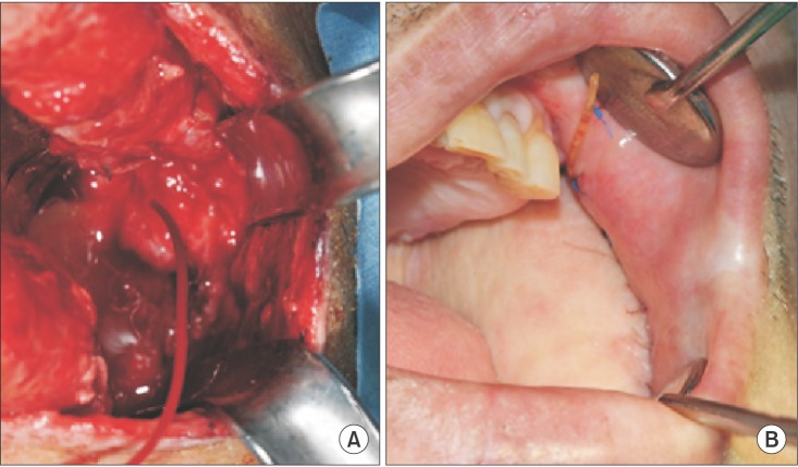
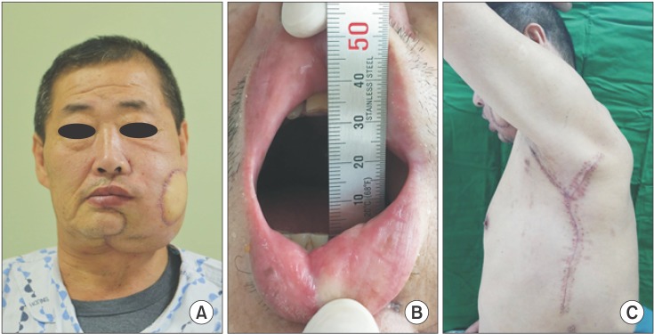
- Squamous cell carcinoma of the buccal mucosa has an aggressive nature, as it grows rapidly and penetrates well with a high recurrence rate. If cancers originating from the buccal mucosa invade adjacent anatomical structures, surgical tumor resection becomes more challenging, thus raising specific considerations for reconstruction relative to the extent of resection. The present case describes the surgical management of a 58-year-old man who presented with persistent ulceration of the mucosal membrane and a mouth-opening limitation of 11 mm. Diagnostic imaging revealed a buccal mucosa tumor that had invaded the retroantral space upward with involvement of the anterior border of the masseter muscle by the lateral part of the tumor. In this report, we present the surgical approach we used to access the masticator space behind the maxillary sinus and discuss how to manage possible damage to Stensen's duct during resection of buccal mucosa tumors.
MeSH Terms
Figure
Reference
-
1. Shah JP, Cendon RA, Farr HW, Strong EW. Carcinoma of the oral cavity. factors affecting treatment failure at the primary site and neck. Am J Surg. 1976; 132:504–507. PMID: 1015542.2. Vegers JW, Snow GB, van der Waal I. Squamous cell carcinoma of the buccal mucosa. A review of 85 cases. Arch Otolaryngol. 1979; 105:192–195. PMID: 426706.
Article3. Kuk SK, Kim BK, Yoon HJ, Hong SD, Hong SP, Lee JI. Investigation on the age and location of oral squamous cell carcinoma incidence in Korea. Korean J Oral Maxillofac Pathol. 2015; 39:393–402.
Article4. Lin CS, Jen YM, Cheng MF, Lin YS, Su WF, Hwang JM, et al. Squamous cell carcinoma of the buccal mucosa: an aggressive cancer requiring multimodality treatment. Head Neck. 2006; 28:150–157. PMID: 16200628.
Article5. Bloom ND, Spiro RH. Carcinoma of the cheek mucosa. A retrospective analysis. Am J Surg. 1980; 140:556–559. PMID: 7425239.6. Conley J, Sadoyama JA. Squamous cell cancer of the buccal mucosa. A review of 90 cases. Arch Otolaryngol. 1973; 97:330–333. PMID: 4573067.
Article7. Lapeyre M, Peiffert D, Malissard L, Hoffstetter S, Pernot M. An original technique of brachytherapy in the treatment of epidermoid carcinomas of the buccal mucosa. Int J Radiat Oncol Biol Phys. 1995; 33:447–454. PMID: 7673032.
Article8. Pop LA, Eijkenboom WM, de Boer MF, de Jong PC, Knegt P, Levendag PC, et al. Evaluation of treatment results of squamous cell carcinoma of the buccal mucosa. Int J Radiat Oncol Biol Phys. 1989; 16:483–487. PMID: 2921152.
Article9. Strome SE, To W, Strawderman M, Gersten K, Devaney KO, Bradford CR, et al. Squamous cell carcinoma of the buccal mucosa. Otolaryngol Head Neck Surg. 1999; 120:375–379. PMID: 10064641.
Article10. Urist MM, O'Brien CJ, Soong SJ, Visscher DW, Maddox WA. Squamous cell carcinoma of the buccal mucosa: analysis of prognostic factors. Am J Surg. 1987; 154:411–414. PMID: 3661845.
Article11. Hakeem AH, Pradhan SA, Tubachi J, Kannan R. Outcome of per oral wide excision of T1-2 N0 localized squamous cell cancer of the buccal mucosa--analysis of 156 cases. Laryngoscope. 2013; 123:177–180. PMID: 22952001.
Article12. Sieczka E, Datta R, Singh A, Loree T, Rigual N, Orner J, et al. Cancer of the buccal mucosa: are margins and T-stage accurate predictors of local control? Am J Otolaryngol. 2001; 22:395–399. PMID: 11713724.
Article13. Castro J, Likhterov I, Mehra S, Bassiri-Tehrani M, Scherl S, Clain J, et al. Approach to en bloc resection and reconstruction of primary masticator space malignancies. Laryngoscope. 2016; 126:372–377. PMID: 26526821.
Article14. Pogrel MA, Kaplan MJ. Surgical approach to the pterygomaxillary region. J Oral Maxillofac Surg. 1986; 44:183–187. PMID: 3456438.
Article15. Dingman DL, Conley J. Lateral approach to the pterygomaxillary region. Ann Otol Rhinol Laryngol. 1970; 79:967–969. PMID: 5506040.
Article16. Spiro RH, Gerold FP, Strong EW. Mandibular "swing" approach for oral and oropharyngeal tumors. Head Neck Surg. 1981; 3:371–378. PMID: 6263826.
Article17. Welvaart K, Caspers RJ, Verkes RJ, Hermans J. The choice between surgical resection and radiation therapy for patients with cancer of the esophagus and cardia: a retrospective comparison between two treatments. J Surg Oncol. 1991; 47:225–229. PMID: 1713631.
Article18. Hiraki A, Yamamoto T, Yoshida R, Nagata M, Kawahara K, Nakagawa Y, et al. Factors affecting volume change of myocutaneous flaps in oral cancer. Int J Oral Maxillofac Surg. 2016; 45:1395–1399. PMID: 27170618.
Article19. Li BH, Jung HJ, Choi SW, Kim SM, Kim MJ, Lee JH. Latissimus dorsi (LD) free flap and reconstruction plate used for extensive maxillo-mandibular reconstruction after tumour ablation. J Craniomaxillofac Surg. 2012; 40:e293–e300. PMID: 22377010.
Article20. Yang ZH, Zhang DM, Chen WL, Wang YY, Fan S. Reconstruction of through-and-through oral cavity defects with folded extended vertical lower trapezius island myocutaneous flap. Br J Oral Maxillofac Surg. 2013; 51:731–735. PMID: 24090763.
Article21. Haggerty CJ, Laughlin RM. Atlas of operative oral and maxillofacial surgery. Ames: Wiley-Blackwell Publishing;2015.22. Van Sickels JE. Management of parotid gland and duct injuries. Oral Maxillofac Surg Clin North Am. 2009; 21:243–246. PMID: 19348990.
Article23. Mehta S, Agrawal J, Dewan AK, Pradhan T. Parotid duct relocation in buccal mucosa cancer resection. J Craniofac Surg. 2014; 25:1746–1747. PMID: 25162543.
Article24. Deygles C Jr, Medeiros R, Carvalho EJ, Carvalho AA. Catheterization of Stenon's duct for surgical excision of oral fibroepithelial hyperplasia. Braz J Otorhinolaryngol. 2012; 78:141. PMID: 22392253.
- Full Text Links
- Actions
-
Cited
- CITED
-
- Close
- Share
- Similar articles
-
- Diagnostic problem of squamous papilloma and oral mucosa malignancy
- VERRUCOUS CARCINOMA A CASE REPORT
- Expression of Cyclooxygenase 1 and 2 in Laryngeal Squamous Cell Carcinoma
- Squamous Cell Carcinoma of the Suprapubic Cystostomy Tract With Bladder Involvement
- Basaloid-Squamous Carcinoma of the Esophagus: A case report


