J Korean Assoc Oral Maxillofac Surg.
2017 Jun;43(3):178-181. 10.5125/jkaoms.2017.43.3.178.
Peripheral odontogenic myxoma in a 12-year-old girl: a rare entity
- Affiliations
-
- 1Department of Oral Pathology and Microbiology, Bharati Vidyapeeth Deemed University Dental College and Hospital, Sangli, India. kmamata@yahoo.com
- 2Department of Pedodontics, Bharati Vidyapeeth Deemed University Dental College and Hospital, Sangli, India.
- KMID: 2386363
- DOI: http://doi.org/10.5125/jkaoms.2017.43.3.178
Abstract
- Peripheral odontogenic myxoma is a rare odontogenic tumor representing an extra osseous counterpart of central odontogenic myxoma. It is commonly seen in gingiva between the 3rd and 4th decades of life and appears predominantly in females. Compared to central odontogenic myxoma, it is a less aggressive, slow-growing lesion with a low recurrence rate. However, close postoperative follow-up is required because of the unlimited growth potential of incompletely removed lesions. It shares many features with other soft tissue myxoid proliferations occurring in the oral cavity and hence needs to be differentiated from them. Very few cases of peripheral odontogenic myxomas have been reported and, to the best of our knowledge, no case has been reported in a pediatric patient. We present an unusual case of peripheral odontogenic myxoma occurring in a 12-year-old girl located in the anterior mandibular gingiva, with an emphasis on differential diagnosis.
Keyword
MeSH Terms
Figure
-
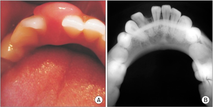
Fig. 1 A. Clinical photograph showing the gingival mass extending buccolingually between teeth #31 and #32. B. Occlusal radiograph showing drifting of #31 and #32 without bone involvement.
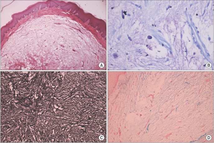
Fig. 2 2. A. H&E stained section (×10) showing loose myxomatous lesional tissue separated from the overlying stratified epithelium by a fibrous capsule. B. Toluidine blue-stained section (×40) showing mast cells in myxoid stroma. C. Reticulin-stained section (×10) showing strong positivity. D. Lesional tissue showing reactivity to Alcian blue staining (×10).
Reference
-
1. Brannon RB. Central odontogenic fibroma, myxoma (odontogenic myxoma, fibromyxoma), and central odontogenic granular cell tumor. Oral Maxillofac Surg Clin North Am. 2004; 16:359–374. PMID: 18088737.
Article2. Jain VK, Reddy SN. Peripheral odontogenic myxoma of maxillary gingiva: a rare clinical entity. J Indian Soc Periodontol. 2013; 17:653–656. PMID: 24174762.
Article3. Reichart PA, Philipsen HP. Odontogenic tumors and allied lesions. London: Quintessence Publishing;2004. p. 189–196.4. Raubenheimer EJ, Noffke CE. Peripheral odontogenic myxoma: a review of the literature and report of two cases. J Maxillofac Oral Surg. 2012; 11:101–104. PMID: 23449257.
Article5. Aytac-Yazicioglu D, Eren H, Görgün S. Peripheral odontogenic myxoma located on the maxillary gingiva: report of a case and review of the literature. Oral Maxillofac Surg. 2008; 12:167–171. PMID: 18642035.
Article6. Mehendiratta M, Rehani S, Solomon MC. The histological spectrum of myxoma, myxofibroma/fibromyxoma and odontogenic fibroma: “a chicken and egg situation”. IOSR J Dent Med Sci. 2012; 1:3–5.7. Perrotti V, Rubini C, Fioroni M, Piattelli A. Soft tissue myxoma: report of an unusual case located on the gingiva. J Clin Periodontol. 2006; 33:76–78. PMID: 16367860.
Article8. Shimoyama T, Horie N, Kato T, Tojo T, Nasu D, Kaneko T, et al. Soft tissue myxoma of the gingiva: report of a case and review of the literature of soft tissue myxoma in the oral region. J Oral Sci. 2000; 42:107–109. PMID: 10989594.
Article9. Whitt J, Barker B, Cobb C. Peripheral odontogenic myxoma. Oral Surg Oral Med Oral Pathol Oral Radiol Endod. 2005; 100:187–188.
Article10. Ramraj PN, Sah SP. Myxoma of oral soft tissue. J Nepal Med Assoc. 2001; 40:274–276.
Article11. Regezi JA, Sciubba JJ, Jordan RCK. Oral pathology: clinical pathologic correlations. 5th ed. St. Louis: Saunders Elsevier;2008. p. 272–273.12. de Assis Caldas Pereira F, Gurgel CA, Ramos EA, Vidal MT, Pinheiro AL, Jurisic V, et al. Distribution of mast cells in benign odontogenic tumors. Tumour Biol. 2012; 33:455–461. PMID: 22125027.13. Mosqueda-Taylor A. New findings and controversies in odontogenic tumors. Med Oral Patol Oral Cir Bucal. 2008; 13:E555–E558. PMID: 18758398.14. Martínez-Mata G, Mosqueda-Taylor A, Carlos-Bregni R, de Almeida OP, Contreras-Vidaurre E, Vargas PA, et al. Odontogenic myxoma: clinico-pathological, immunohistochemical and ultrastructural findings of a multicentric series. Oral Oncol. 2008; 44:601–607. PMID: 17996487.
Article15. Barnes L, Eveson JW, Reichart P, Sidransky D. World Health Organization classification of tumors. Pathology and genetics of head and neck tumours. Lyon: IARC Press;2005. p. 197.16. Choi SH, Jeong JC, Song MS, Seo JH, Kim SB, Jun CH. A case of odontogenic myxoma related to both impacted canine teeth in the mandible. J Korean Assoc Oral Maxillofac Surg. 2003; 29:64–67.
- Full Text Links
-
- Actions
-
Cited
- CITED
-
- Close
- Share
-
- Similar articles
-
- Coexistence of Odontogenic Myxoma and Dentigerous Cyst on Mandible: A Case Report
- Fabrication of complete dentures for a patient with odontogenic myxoma: A case report
- Odontogenic myxoma of the mandible: Report of a case
- A case of odontogenic myxoma related to both impacted canine teeth in the mandible
- Central odontogenic fibroma: a case report
