Extraforaminal Extrusion of Intervertebral Disc Misdiagnosed as Neurogenic Tumor: a Case Report
- Affiliations
-
- 1Department of Rehabilitation Medicine, Gyeongsang National University School of Medicine and Gyeongsang National University Changwon Hospital, Changwon, Korea.
- 2Department of Neurosurgery, Gyeongsang National University School of Medicine and Gyeongsang National University Changwon Hospital, Changwon, Korea.
- 33Department of Radiology, Gyeongsang National University School of Medicine and Gyeongsang National University Changwon Hospital, Changwon, Korea. drlotus@naver.com
- KMID: 2385609
- DOI: http://doi.org/10.13104/imri.2017.21.2.109
Abstract
- A 55-year-old male presented with dysesthesia of the right anteromedial thigh. A magnetic resonance image revealed a globular mass at the right extraforaminal area of the L3/4 level. Based on the diagnosis of neurogenic tumor, surgical excision was performed. The surgical impression and pathology confirmed extrusion of intervertebral disc. In a retrospective review of the magnetic resonance image, we noticed a fibrillary pattern directed from the intervertebral disc space to the lesion, and disrupted annulus fibrosus and indentation caused by the ring apophysis. We suggest aforementioned features, indicative of intervertebral disc lesions, to be checked when interpreting mass lesions around the intervertebral foramen.
Keyword
MeSH Terms
Figure
-
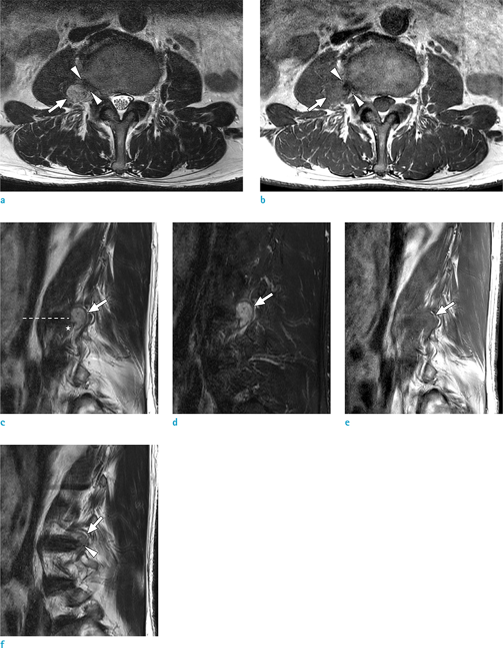
Fig. 1 Axial (a) and sagittal (c) T2-weighted images of the L3/4 disc level show a hyperintense mass (arrows) with a dark signal rim at the right extraforaminal zone. There is a fibrillary pattern originating from the intervertebral disc to the mass (arrowheads in a). Indentation of the mass (star in c) is observed just below the intervertebral disc level (dotted line). A sagittal T2-weighted image obtained at the medial aspect of the mass (f) shows continuity (arrowhead) between the disc and the mass (arrow) with disruption of the annulus fibrosus. Axial (b) and sagittal (e) T1-weighted images show isointensity of the mass (arrow) and hypointense lesion (arrowheads) corresponding to fibrillary pattern in a. Fat-suppressed sagittal T2-weighted image (d) shows hyperintensity of the mass (arrow).

Fig. 2 Intraoperative photograph (a) shows the mass (white arrow) that is continuous with the disc space. The nerve root (black arrow) is separate from the mass. Photograph after excision of the mass (b) shows an empty cavity of intervertebral disc space (arrow).
Reference
-
1. Bakar B, Sumer MM, Cila A, Tekkok IH. An extreme lateral lumbar disc herniation mimicking L4 schwannoma. Acta Neurol Belg. 2009; 109:155–158.2. Dimogerontas G, Paidakakos NA, Konstantinidis E. Voluminous free disk fragment mimicking an extradural tumor. Neurol Med Chir (Tokyo). 2012; 52:656–665.3. Fardon DF, Williams AL, Dohring EJ, Murtagh FR, Gabriel Rothman SL, Sze GK. Lumbar disc nomenclature: version 2.0: Recommendations of the combined task forces of the North American Spine Society, the American Society of Spine Radiology and the American Society of Neuroradiology. Spine J. 2014; 14:2525–2545.4. Bloomer CW, Ackerman A, Bhatia RG. Imaging for spine tumors and new applications. Top Magn Reson Imaging. 2006; 17:69–87.5. Abreu E, Aubert S, Wavreille G, Gheno R, Canella C, Cotten A. Peripheral tumor and tumor-like neurogenic lesions. Eur J Radiol. 2013; 82:38–50.6. Jee WH, Oh SN, McCauley T, et al. Extraaxial neurofibromas versus neurilemmomas: discrimination with MRI. AJR Am J Roentgenol. 2004; 183:629–633.7. Abul-Kasim K, Thurnher MM, McKeever P, Sundgren PC. Intradural spinal tumors: current classification and MRI features. Neuroradiology. 2008; 50:301–314.8. Pilavaki M, Chourmouzi D, Kiziridou A, Skordalaki A, Zarampoukas T, Drevelengas A. Imaging of peripheral nerve sheath tumors with pathologic correlation: pictorial review. Eur J Radiol. 2004; 52:229–239.9. Weyreuther M, Heyde CE, Westphal M, Zierski J, Weber U. MRI atlas. Orthopedics and neurosurgery, the spine. Berlin, Heidelberg: Springer-Verlag;2007. p. 106–107.10. Daghighi MH, Pouriesa M, Maleki M, et al. Migration patterns of herniated disc fragments: a study on 1,020 patients with extruded lumbar disc herniation. Spine J. 2014; 14:1970–1977.
- Full Text Links
-
- Actions
-
Cited
- CITED
-
- Close
- Share
-
- Similar articles
-
- Far lateral lumbar disc extrusion in a dachshund dog
- Paramedian Tangential Approach for the Lumbosacral Extraforaminal Disc Herniations
- Spontaneous Regression of a Large Lumbar Disc Extrusion
- The Morphometric Analysis of the Extraforamen in the Lumbosacral Spine: Magnetic Resonance Imaging and Computed Tomography Study
- Correlation of Magnetic Resonance Imaging of Lumbar Herniated Intervertebral Disc with Operative Findings
