J Korean Soc Radiol.
2017 Jul;77(1):53-56. 10.3348/jksr.2017.77.1.53.
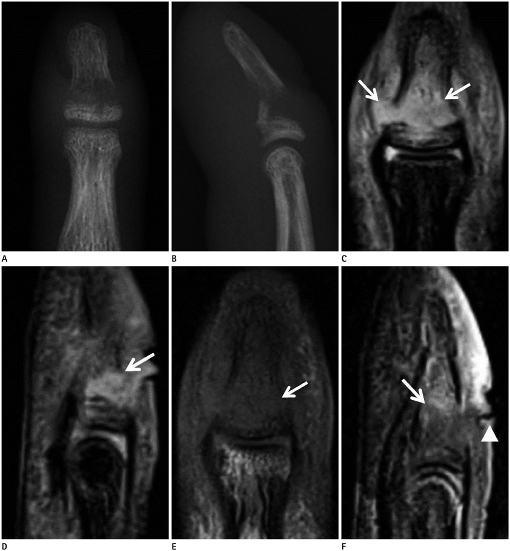
MRI Findings of Post-Traumatic Osteomyelitis of Distal Phalanx Following Neglected Open Fracture
- Affiliations
-
- 1Department of Radiology, Inje University Sanggye Paik Hospital, Seoul, Korea. merita@paik.ac.kr
- KMID: 2384736
- DOI: http://doi.org/10.3348/jksr.2017.77.1.53
Abstract
- Careful radiologic examination of the osteolytic lesion is important for patients with fracture. Differential diagnosis includes osteonecrosis, neoplasm and infections. In this report, we presented MRI findings of post-traumatic osteomyelitis following neglected open fracture of 3rd distal phalanx with open wound. Early suspicion and imaging of wound or soft tissue inflammation around osteolytic lesion could be helpful for diagnosis of osteomyelitis.
MeSH Terms
Figure
Reference
-
1. Roesgen M, Hierholzer G, Hax PM. Post-traumatic osteomyelitis. Pathophysiology and management. Arch Orthop Trauma Surg. 1989; 108:1–9.2. Shimose S, Sugita T, Kubo T, Matsuo T, Nobuto H, Ochi M. Differential diagnosis between osteomyelitis and bone tumors. Acta Radiol. 2008; 49:928–933.3. Balakrishnan C, Vashi C, Jackson O, Hess J. Post-traumatic osteomyelitis of the clavicle: a case report and review of literature. Can J Plast Surg. 2008; 16:89–91.4. Pineda C, Espinosa R, Pena A. Radiographic imaging in osteomyelitis: the role of plain radiography, computed tomography, ultrasonography, magnetic resonance imaging, and scintigraphy. Semin Plast Surg. 2009; 23:80–89.5. Gold RH, Hawkins RA, Katz RD. Bacterial osteomyelitis: findings on plain radiography, CT, MR, and scintigraphy. AJR Am J Roentgenol. 1991; 157:365–370.6. Capparelli G, Barresi D, Bertucci B, Stanà C, Cristofaro A, Tamburrini S. [Magnetic resonance findings in chronic osteomyelitis fistula]. Radiol Med. 1998; 96:434–438.
- Full Text Links
- Actions
-
Cited
- CITED
-
- Close
- Share
- Similar articles
-
- A Case of Digital Intraosseous Epidermoid Inclusion Cyst of the Distal Phalanx
- Intraosseous Epidermoid Cyst of Distal Phalanx: A case report
- Multiple Pinning in Base Fracture of Distal Phalanx
- Treatement of Osteomyelitis and Soft Tissue Infection of Fingers Using Mini External Fixator
- Neglected Traumatic Posterior Hip Dislocation in a Crutch-walking Patient: A Case Report


