Application of augmented reality for inferior alveolar nerve block anesthesia: A technical note
- Affiliations
-
- 1Department of Oral and Maxillofacial Surgery, National Health Insurance Service Ilsan Hospital, Goyang, Republic of Korea. omskang@nhimc.or.kr
- KMID: 2384474
- DOI: http://doi.org/10.17245/jdapm.2017.17.2.129
Abstract
- Efforts to apply augmented reality (AR) technology in the medical field include the introduction of AR techniques into dental practice. The present report introduces a simple method of applying AR during an inferior alveolar nerve block, a procedure commonly performed in dental clinics.
Figure
-

Fig. 1 A 3-dimensional reconstruction of the mandible generated from computed tomography images using the 3-dimensional simulation software.
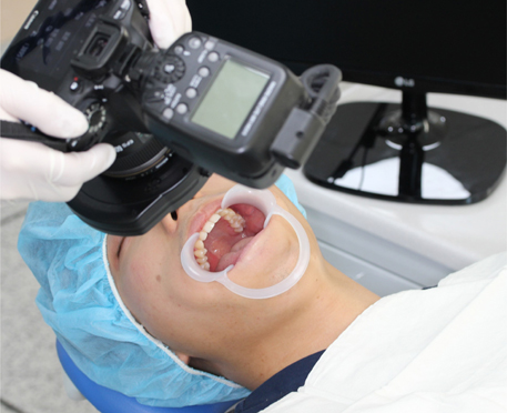
Fig. 2 Intraoral photographs were taken from the angle at which the clinician used to look down at the patient's oral cavity for a right-sided inferior alveolar nerve block anesthesia.
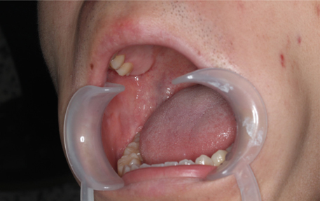
Fig. 3 Intraoral photographs were taken from the angle at which the clinician used to look down at the patient's oral cavity for injection of local anesthetics for right side inferior alveolar nerve block anesthesia.
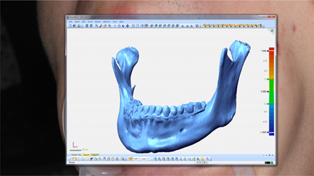
Fig. 4 The window of the 3-dimensional simulation software with the 3-dimensional mandibular image overlapping the intraoral photograph.
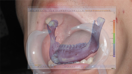
Fig. 5 Adjustment of the window transparency of the 3-dimensional simulation software to enable visualization of the intraoral photograph through the 3-dimensional mandibular image.

Fig. 6 Overlapping of the 3-dimensional mandibular images from the simulation software onto the intraoral photographs by matching positions of the teeth between the 3-dimensional mandibular images and intraoral photographs.
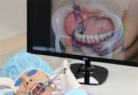
Fig. 7 Performing of an inferior alveolar nerve block procedure using a simple augmented reality method, in which the superimposed images are used as references to locate the mandibular foramen in an intraoral view during the injection of local anesthetics.
Cited by 1 articles
-
Facial blanching after inferior alveolar nerve block anesthesia: an unusual complication
Sang-Hoon Kang, Yu-Jin Won
J Dent Anesth Pain Med. 2017;17(4):317-321. doi: 10.17245/jdapm.2017.17.4.317.
Reference
-
1. Suenaga H, Tran HH, Liao H, Masamune K, Dohi T, Hoshi K, et al. Vision-based markerless registration using stereo vision and an augmented reality surgical navigation system: a pilot study. BMC Med Imaging. 2015; 15:51.
Article2. Zhu M, Liu F, Chai G, Pan JJ, Jiang T, Lin L, et al. A novel augmented reality system for displaying inferior alveolar nerve bundles in maxillofacial surgery. Sci Rep. 2017; 7:42365.
Article3. Wang J, Suenaga H, Hoshi K, Yang L, Kobayashi E, Sakuma I, et al. Augmented reality navigation with automatic marker-free image registration using 3-D image overlay for dental surgery. IEEE Trans Biomed Eng. 2014; 61:1295–1304.
Article4. Hernandez Y, Tarazona B, Zamora N, Cibrian R, Gandia J, Paredes V. Comparative study of reproducibility and accuracy in measuring mesiodistal tooth sizes using three different methods: 2D digital, 3D CBCT, and 3D CBCT segmented. Oral Radiol. 2015; 31:165–172.
Article5. Badiali G, Ferrari V, Cutolo F, Freschi C, Caramella D, Bianchi A, et al. Augmented reality as an aid in maxillofacial surgery: validation of a wearable system allowing maxillary repositioning. J Craniomaxillofac Surg. 2014; 42:1970–1976.
Article6. Kang SH, Lee JW, Lim SH, Kim YH, Kim MK. Dental image replacement on cone beam computed tomography with three-dimensional optical scanning of a dental cast, occlusal bite, or bite tray impression. Int J Oral Maxillofac Surg. 2014; 43:1293–1301.
Article7. You TM, Kim KD, Huh J, Woo EJ, Park W. The influence of mandibular skeletal characteristics on inferior alveolar nerve block anesthesia. J Dent Anesth Pain Med. 2015; 15:113–119.
Article8. Kang SH, Byun IY, Kim JH, Park HK, Kim MK. Three-dimensional anatomic analysis of mandibular foramen with mandibular anatomic landmarks for inferior alveolar nerve block anesthesia. Oral Surg Oral Med Oral Pathol Oral Radiol. 2013; 115:e17–e23.
Article9. Wang J, Suenaga H, Liao H, Hoshi K, Yang L, Kobayashi E, et al. Real-time computer-generated integral imaging and 3D image calibration for augmented reality surgical navigation. Comput Med Imaging Graph. 2015; 40:147–159.
Article10. Suenaga H, Hoang Tran H, Liao H, Masamune K, Dohi T, Hoshi K, et al. Real-time in situ three-dimensional integral videography and surgical navigation using augmented reality: a pilot study. Int J Oral Sci. 2013; 5:98–102.
Article
- Full Text Links
-
- Actions
-
Cited
- CITED
-
- Close
- Share
-
- Similar articles
-
- Updates on the Inferior Alveolar Nerve Block Anesthesia
- Diplopia after Inferior Alveolar Nerve Block Anesthesia: A Case Report
- Facial blanching after inferior alveolar nerve block anesthesia: an unusual complication
- Diplopia after inferior alveolar nerve block: case report and related physiology
- Transient Visual Acuity Decrease after Inferior Alveolar Nerve Block Anesthesia
