Three-dimensional measurement of periodontal surface area for quantifying inflammatory burden
- Affiliations
-
- 1Department of Oral Anatomy, Dankook University College of Dentistry, Cheonan, Korea. pjtdent@gmail.com
- 2Department of Pediatric Dentistry, Wonkwang University, Daejeon, Korea.
- 3Department of Dentomaxillofacial Radiology, Dankook University College of Dentistry, Cheonan, Korea.
- KMID: 2383829
- DOI: http://doi.org/10.5051/jpis.2017.47.3.154
Abstract
- PURPOSE
Measurement of the root surface area (RSA) is important in periodontal treatment and for the evaluation of periodontal disease as a risk factor for systemic disease. The aim of this study was to measure the RSA at 6 mm below the cementoenamel junction (CEJ) using the Mimics software (Materialise, Leuven, Belgium).
METHODS
We obtained cone-beam computed tomography (CBCT) data from 33 patients who had visited the Department of Oral and Maxillofacial Radiology of Dankook University Dental Hospital. The patients comprised 17 men and 16 women aged from 20 to 35 years, with a mean age of 24.4 years. Only morphologically intact teeth were included in our data. Because the third molars of the maxilla and mandible have a high deformation rate and were absent in some participants, they were not included in our research material.
RESULTS
The CBCT data were reconstructed into 3-dimensional (3D) teeth models using the Mimics software, and the RSA at 6 mm below the CEJ was separated and measured using 3-Matic (Materialise). In total, 924 3D teeth models were created, and the area at 6 mm below the CEJ could be isolated in all the models. The area at 6 mm below the CEJ was measured in all teeth from the 33 patients and compared based on sex and position (maxilla vs. mandible).
CONCLUSIONS
In this study, we demonstrated that it was feasible to generate 3D data and to evaluate RSA values using CBCT and the Mimics software. These results provide deeper insights into the relationship between periodontal inflammatory burden and systemic diseases.
MeSH Terms
Figure
-

Figure 1 Process of masking mandibular teeth in CBCT. (A) Thresholding mask based on the general bone value in Mimics (Materialise, Leuven, Belgium). (B) Separating soft tissue and cervical bone in the mandible, and then filling inside the cavity. (C) Extracting teeth from the mandible.CBCT: cone-beam computed tomography.
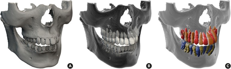
Figure 2 Process of generating the 3D structure of teeth. (A) The 3D skull model was generated using Mimics (Materialise, Leuven, Belgium). (B) The skull and teeth were separated using the region-growing method. (C) The teeth are indicated in red and orange in the maxilla, and green and blue in the mandible. The teeth were extracted and bones were deleted.3D: 3-dimensional.

Figure 3 Evaluation of the CAL in periodontitis. The CEJ is the cervical line between the crown and root. The CAL is the distance from the attachment level to the CEJ. The PPD is the distance from the attachment level to the gingival margin.CAL: clinical attachment level, CEJ: cementoenamel junction, PPD: probing pocket depth.
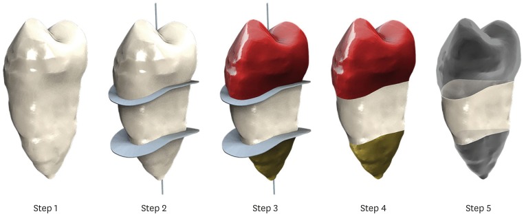
Figure 4 Separation of the RSA at 6 mm below the CEJ. Step 1. Generated using 3-dimensional data from CBCT. Step 2. A root line position is created around the tooth meridian and a cross section of the root at 6 mm below it. Step 3. Teeth are divided into 3 parts based on 2 apical surfaces. Step 4. The root cross section is removed. Step 5. The lower part and crown are removed, except the area 6 mm below the CEJ.RSA: root surface area, CEJ: cementoenamel junction, CBCT: cone-beam computed tomography.
Reference
-
1. Offenbacher S, Beck JD. Commentary: changing paradigms in the oral disease-systemic disease relationship. J Periodontol. 2014; 85:761–764. PMID: 24875011.
Article2. Beck J, Garcia R, Heiss G, Vokonas PS, Offenbacher S. Periodontal disease and cardiovascular disease. J Periodontol. 1996; 67:1123–1137. PMID: 8910831.
Article3. Offenbacher S, Katz V, Fertik G, Collins J, Boyd D, Maynor G, et al. Periodontal infection as a possible risk factor for preterm low birth weight. J Periodontol. 1996; 67:1103–1113. PMID: 8910829.
Article4. Chistiakov DA, Orekhov AN, Bobryshev YV. Links between atherosclerotic and periodontal disease. Exp Mol Pathol. 2016; 100:220–235. PMID: 26777261.
Article5. Haynes WG, Stanford C. Periodontal disease and atherosclerosis: from dental to arterial plaque. Arterioscler Thromb Vasc Biol. 2003; 23:1309–1311. PMID: 12909566.6. Haraszthy VI, Zambon JJ, Trevisan M, Zeid M, Genco RJ. Identification of periodontal pathogens in atheromatous plaques. J Periodontol. 2000; 71:1554–1560. PMID: 11063387.
Article7. Górska R, Gregorek H, Kowalski J, Laskus-Perendyk A, Syczewska M, Madaliński K. Relationship between clinical parameters and cytokine profiles in inflamed gingival tissue and serum samples from patients with chronic periodontitis. J Clin Periodontol. 2003; 30:1046–1052. PMID: 15002890.
Article8. Leibur E, Tuhkanen A, Pintson U, Söder PO. Prostaglandin E2 levels in blood plasma and in crevicular fluid of advanced periodontitis patients before and after surgical therapy. Oral Dis. 1999; 5:223–228. PMID: 10483068.
Article9. Bansal J, Bansal A, Kukreja N, Kukreja U. Periodontal diseases as an emerging potential risk factor for adverse pregnancy outcomes: a review of concepts. J Turk Ger Gynecol Assoc. 2011; 12:176–180. PMID: 24591987.
Article10. Hujoel PP, White BA, García RI, Listgarten MA. The dentogingival epithelial surface area revisited. J Periodontal Res. 2001; 36:48–55. PMID: 11246704.
Article11. Yamamoto T, Kinoshita Y, Tsuneishi M, Takizawa H, Umemura O, Watanabe T. Estimation of the remaining periodontal ligament from attachment-level measurements. J Clin Periodontol. 2006; 33:221–225. PMID: 16489949.
Article12. Nesse W, Abbas F, van der Ploeg I, Spijkervet FK, Dijkstra PU, Vissink A. Periodontal inflamed surface area: quantifying inflammatory burden. J Clin Periodontol. 2008; 35:668–673. PMID: 18564145.
Article13. Gu Y, Tang Y, Zhu Q, Feng X. Measurement of root surface area of permanent teeth with root variations in a Chinese population-A micro-CT analysis. Arch Oral Biol. 2016; 63:75–81. PMID: 26723016.
Article14. Choi JW, Kim N. Clinical application of three-dimensional printing technology in craniofacial plastic surgery. Arch Plast Surg. 2015; 42:267–277. PMID: 26015880.
Article15. Mattila K, Rasi V, Nieminen M, Valtonen V, Kesäniemi A, Syrjälä S, et al. von Willebrand factor antigen and dental infections. Thromb Res. 1989; 56:325–329. PMID: 2617473.
Article16. Syrjänen J, Peltola J, Valtonen V, Iivanainen M, Kaste M, Huttunen JK. Dental infections in association with cerebral infarction in young and middle-aged men. J Intern Med. 1989; 225:179–184. PMID: 2703800.17. Bahekar AA, Singh S, Saha S, Molnar J, Arora R. The prevalence and incidence of coronary heart disease is significantly increased in periodontitis: a meta-analysis. Am Heart J. 2007; 154:830–837. PMID: 17967586.
Article18. Blaizot A, Vergnes JN, Nuwwareh S, Amar J, Sixou M. Periodontal diseases and cardiovascular events: meta-analysis of observational studies. Int Dent J. 2009; 59:197–209. PMID: 19774803.19. Simpson TC, Weldon JC, Worthington HV, Needleman I, Wild SH, Moles DR, et al. Treatment of periodontal disease for glycaemic control in people with diabetes mellitus. Cochrane Database Syst Rev. 2015; CD004714. PMID: 26545069.
Article20. Teeuw WJ, Gerdes VE, Loos BG. Effect of periodontal treatment on glycemic control of diabetic patients: a systematic review and meta-analysis. Diabetes Care. 2010; 33:421–427. PMID: 20103557.21. Boggess KA, Moss K, Madianos P, Murtha AP, Beck J, Offenbacher S. Fetal immune response to oral pathogens and risk of preterm birth. Am J Obstet Gynecol. 2005; 193:1121–1126. PMID: 16157123.
Article22. Chiu B. Multiple infections in carotid atherosclerotic plaques. Am Heart J. 1999; 138:S534–S536. PMID: 10539867.
Article23. Jared H, Boggess KA, Moss K, Bose C, Auten R, Beck J, et al. Fetal exposure to oral pathogens and subsequent risk for neonatal intensive care admission. J Periodontol. 2009; 80:878–883. PMID: 19485816.
Article24. Mustapha IZ, Debrey S, Oladubu M, Ugarte R. Markers of systemic bacterial exposure in periodontal disease and cardiovascular disease risk: a systematic review and meta-analysis. J Periodontol. 2007; 78:2289–2302. PMID: 18052701.
Article25. Paraskevas S, Huizinga JD, Loos BG. A systematic review and meta-analyses on C-reactive protein in relation to periodontitis. J Clin Periodontol. 2008; 35:277–290. PMID: 18294231.
Article26. Al-Shammari KF, Al-Khabbaz AK, Al-Ansari JM, Neiva R, Wang HL. Risk indicators for tooth loss due to periodontal disease. J Periodontol. 2005; 76:1910–1918. PMID: 16274310.
Article27. Burt B. Research, Science and Therapy Committee of the American Academy of Periodontology. Position paper: epidemiology of periodontal diseases. J Periodontol. 2005; 76:1406–1419. PMID: 16101377.28. Kamburoğlu K, Kolsuz E, Kurt H, Kiliç C, Özen T, Paksoy CS. Accuracy of CBCT measurements of a human skull. J Digit Imaging. 2011; 24:787–793. PMID: 20857166.
Article29. Shen P, Zhao J, Fan L, Qiu H, Xu W, Wang Y, et al. Accuracy evaluation of computer-designed surgical guide template in oral implantology. J Craniomaxillofac Surg. 2015; 43:2189–2194. PMID: 26776292.
Article30. Forst D, Nijjar S, Flores-Mir C, Carey J, Secanell M, Lagravere M. Comparison of in vivo 3D cone-beam computed tomography tooth volume measurement protocols. Prog Orthod. 2014; 15:69. PMID: 25534123.
Article31. Li W, Chen F, Zhang F, Ding W, Ye Q, Shi J, et al. Volumetric measurement of root resorption following molar mini-screw implant intrusion using cone beam computed tomography. PLoS One. 2013; 8:e60962. PMID: 23585866.
Article32. Wang Y, He S, Yu L, Li J, Chen S. Accuracy of volumetric measurement of teeth in vivo based on cone beam computer tomography. Orthod Craniofac Res. 2011; 14:206–212. PMID: 22008300.33. Al-Rawi B, Hassan B, Vandenberge B, Jacobs R. Accuracy assessment of three-dimensional surface reconstructions of teeth from cone beam computed tomography scans. J Oral Rehabil. 2010; 37:352–358. PMID: 20180895.
Article
- Full Text Links
-
- Actions
-
Cited
- CITED
-
- Close
- Share
-
- Similar articles
-
- Three dimensional analysis of Korean dentogingival complex
- Root surface roughness following mechanical instrumentation, in vitro 3 dimensional planimetric study
- The SEM Observation of The Various Root Treatment Effect On Furcation Area
- Comparison of 2 root surface area measurement methods: 3-dimensional laser scanning and cone-beam computed tomography
- Periodontal attachment loss of extracted teeth for periodontal reasons
