Clin Endosc.
2017 Mar;50(2):197-201. 10.5946/ce.2016.085.
Fluoroscopy-Guided Endoscopic Removal of Foreign Bodies
- Affiliations
-
- 1Department of Internal Medicine, Asan Digestive Disease Research Institute, Asan Medical Center, University of Ulsan College of Medicine, Seoul, Korea.
- 2Department of Gastroenterology, Asan Digestive Disease Research Institute, Asan Medical Center, University of Ulsan College of Medicine, Seoul, Korea. ji110@hanmail.net
- KMID: 2383538
- DOI: http://doi.org/10.5946/ce.2016.085
Abstract
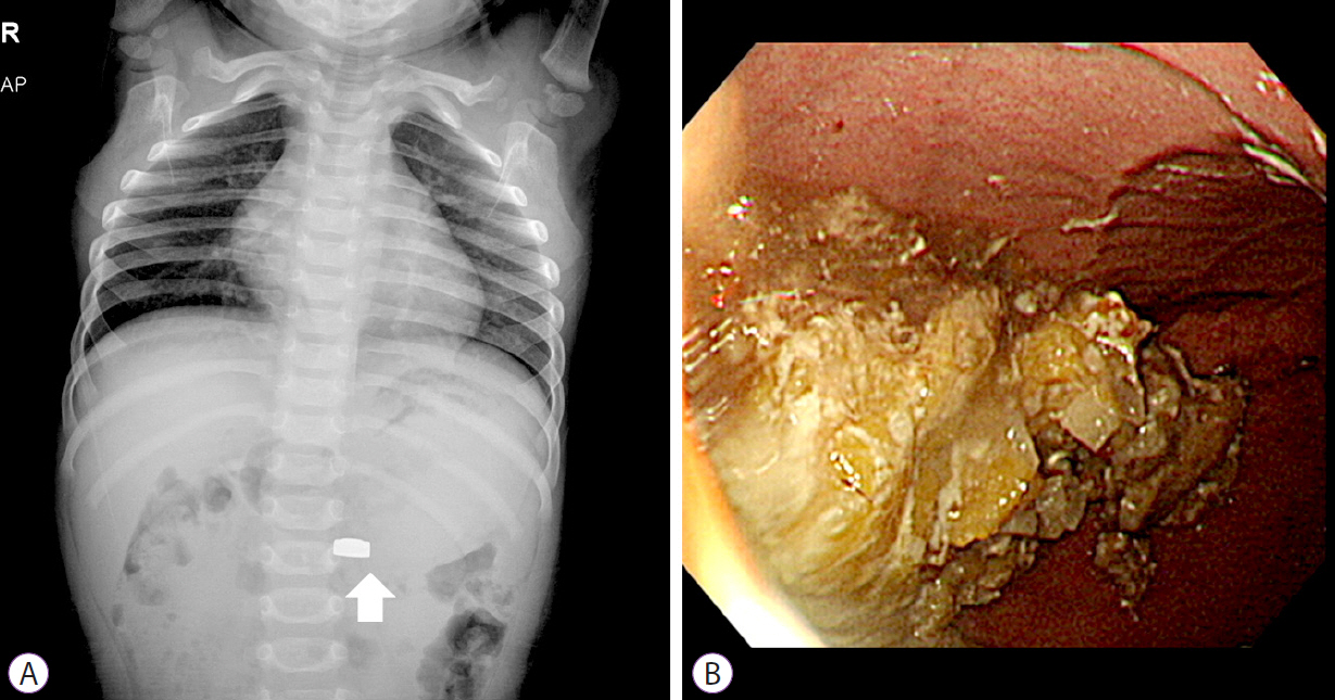
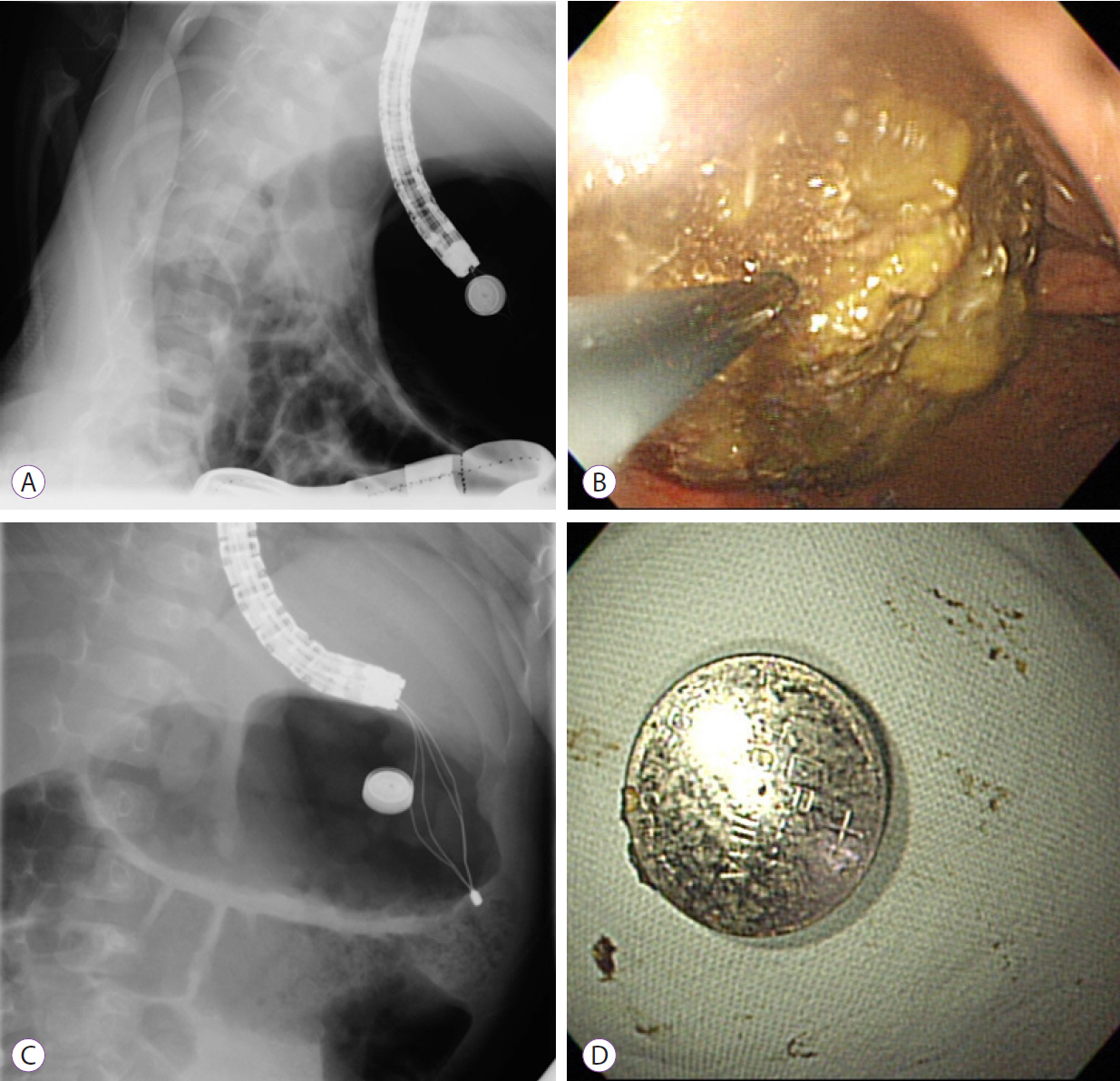
- In most cases of ingested foreign bodies, endoscopy is the first treatment of choice. Moreover, emergency endoscopic removal is required for sharp and pointed foreign bodies such as animal or fish bones, food boluses, and button batteries due to the increased risks of perforation, obstruction, and bleeding. Here, we presented two cases that needed emergency endoscopic removal of foreign bodies without sufficient fasting time. Foreign bodies could not be visualized by endoscopy due to food residue; therefore, fluoroscopic imaging was utilized for endoscopic removal of foreign bodies in both cases.
Figure
Cited by 1 articles
-
섭취 수년 후 발견된 위내 자석 이물
Dong Chan Joo, Moon Won Lee, Seung Min Hong, Dong Hoon Baek, Bong Eun Lee, Gwang Ha Kim, Geun Am Song
Korean J Gastroenterol. 2023;82(4):198-201. doi: 10.4166/kjg.2023.087.
Reference
-
1. Tang SJ. Endoscopic management of foreign bodies in the gastrointestinal tract. Video Journal and Encyclopedia of GI Endoscopy. 2013; 1:35–38.
Article2. ASGE Standards of Practice Committee, Ikenberry SO, Jue TL, et al. Management of ingested foreign bodies and food impactions. Gastrointest Endosc. 2011; 73:1085–1091.
Article3. Sugawa C, Ono H, Taleb M, Lucas CE. Endoscopic management of foreign bodies in the upper gastrointestinal tract: a review. World J Gastrointest Endosc. 2014; 6:475–481.
Article4. Birk M, Bauerfeind P, Deprez PH, et al. Removal of foreign bodies in the upper gastrointestinal tract in adults: European society of gastrointestinal endoscopy (ESGE) clinical guideline. Endoscopy. 2016; 48:489–496.
Article5. Anderson KL, Dean AJ. Foreign bodies in the gastrointestinal tract and anorectal emergencies. Emerg Med Clin North Am. 2011; 29:369–400. ix.
Article6. Lee HJ, Kim HS, Jeon J, et al. Endoscopic foreign body removal in the upper gastrointestinal tract: risk factors predicting conversion to surgery. Surg Endosc. 2016; 30:106–113.
Article7. Hong KH, Kim YJ, Kim JH, Chun SW, Kim HM, Cho JH. Risk factors for complications associated with upper gastrointestinal foreign bodies. World J Gastroenterol. 2015; 21:8125–8131.
Article
- Full Text Links
- Actions
-
Cited
- CITED
-
- Close
- Share
- Similar articles
-
- Removal of Esophageal Foreign Body: Fluoroscopic Guided Double Balloon Technique
- Removal of foreign body on cheek using endoscope and C-arm fluoroscopy
- Endoscopic Removal of Multiple Toothbrushes: A Case Report
- Retrieval of Metallic Foreign Bodies from the Upper Airway Using Intraoperative C-Arm Fluoroscopy: Case Report and Literature Review
- Upper Gastrointestinal Tract Foreign Bodies


