Korean J Gastroenterol.
2016 Dec;68(6):321-325. 10.4166/kjg.2016.68.6.321.
A Case of Hepatocellular Carcinoma Presenting as a Gingival Mass
- Affiliations
-
- 1Department of Internal Medicine, Seoul Paik Hospital, Inje University College of Medicine, Seoul, Korea. rshdrryu@hanmail.net
- 2Department of Pathology, Seoul Paik Hospital, Inje University College of Medicine, Seoul, Korea.
- KMID: 2383463
- DOI: http://doi.org/10.4166/kjg.2016.68.6.321
Abstract
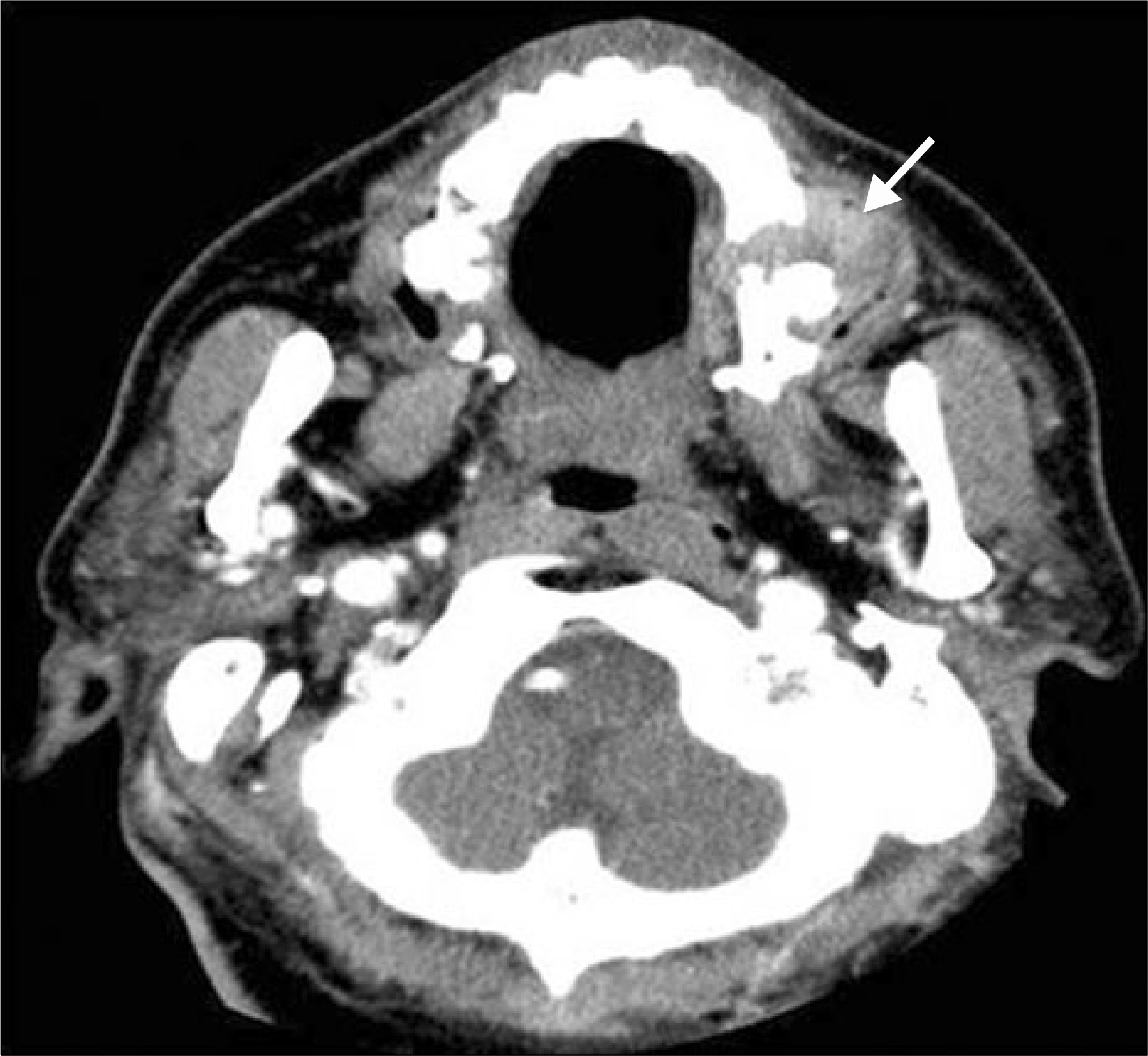
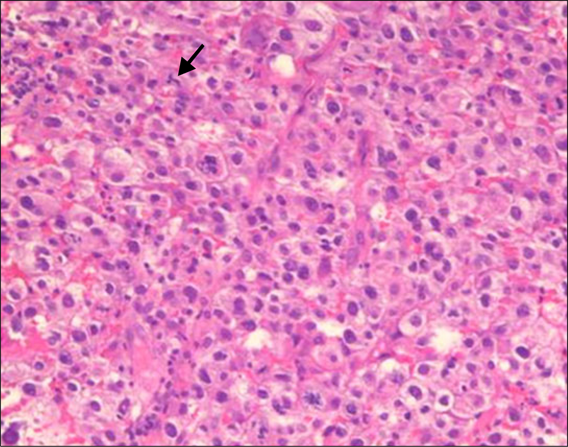
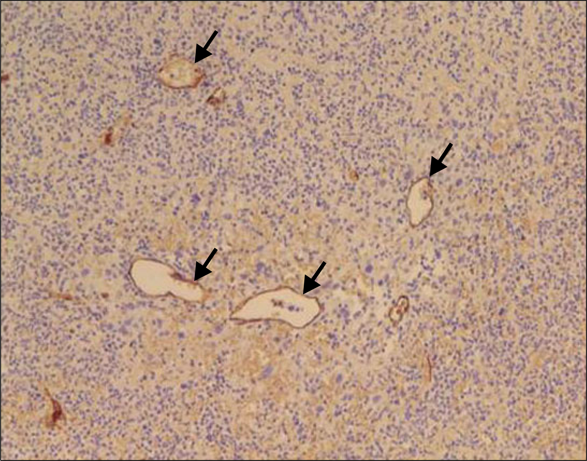
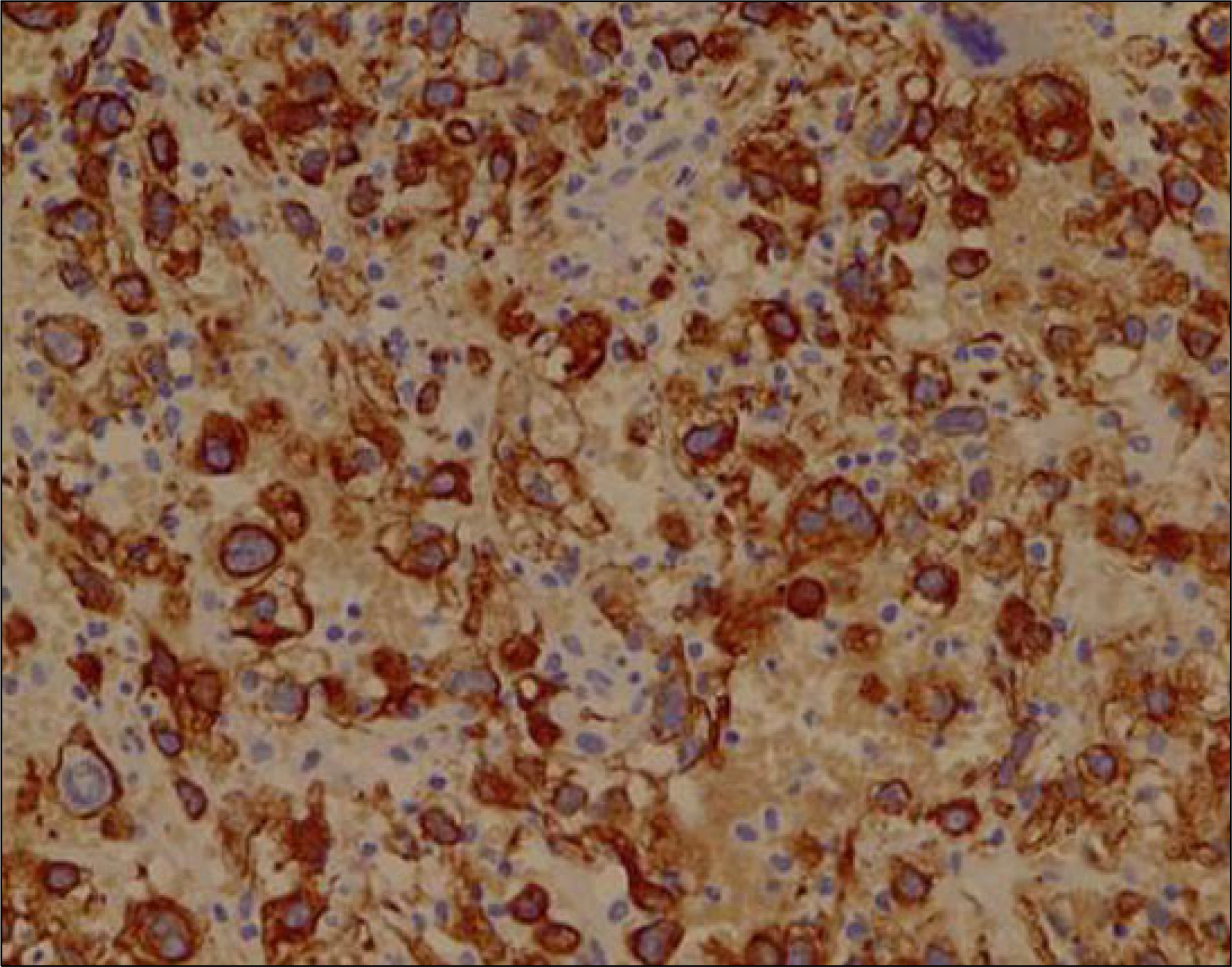
- Oral metastatic tumor, which is uncommon and represents less than 1% of malignant oral neoplasms, usually arises from a primary mucosal or cutaneous cancer located in the head and neck regions. Metastasis of hepatocellular carcinoma (HCC) to the oral cavity, especially to gingiva, is extremely rare. A 50-year-old man, who was a chronic alcoholic and hepatitis B virus carrier, presented with abdominal distension and weight loss for the past 3 months. Three-phased contrast-enhanced abdominal CT revealed numerous conglomerated masses in the liver, suggesting huge HCCs arising in the background of liver cirrhosis with a large amount of ascites. He complained of recurrent profuse bleeding from the left upper gingival mass. A facial CT revealed an oral cavity mass destructing the left maxillary alveolar process and hard palate, which was diagnosed as metastatic HCC by an incisional biopsy. Herein, we report a case of metastatic HCC to the gingiva.
Keyword
MeSH Terms
Figure
Cited by 1 articles
-
Clinicopathologic features of cutaneous metastases from internal malignancies
Hyeong Mok Kwon, Gyu Yeong Kim, Dong Hoon Shin, Young Kyung Bae
J Pathol Transl Med. 2021;55(4):289-297. doi: 10.4132/jptm.2021.05.24.
Reference
-
References
1. Kao JH, Chen DS. Changing disease burden of hepatocellular carcinoma in the Far East and Southeast Asia. Liver Int. 2005; 25:696–703.
Article2. Huang SF, Wu RC, Chang JT, et al. Intractable bleeding from solitary mandibular metastasis of hepatocellular carcinoma. World J Gastroenterol. 2007; 13:4526–4528.
Article3. Sawabe M, Nakamura T, Kanno J, Kasuga T. Analysis of morphological factors of hepatocellular carcinoma in 98 autopsy cases with respect to pulmonary metastasis. Acta Pathol Jpn. 1987; 37:1389–1404.
Article4. Katyal S, Oliver JH 3rd, Peterson MS, Ferris JV, Carr BS, Baron RL. Extrahepatic metastases of hepatocellular carcinoma. Radiology. 2000; 216:698–703.
Article5. Natsuizaka M, Omura T, Akaike T, et al. Clinical features of hepatocellular carcinoma with extrahepatic metastases. J Gastroenterol Hepatol. 2005; 20:1781–1787.
Article6. Watanabe J, Nakashima O, Kojiro M. Clinicopathologic study on lymph node metastasis of hepatocellular carcinoma: a retrospective study of 660 consecutive autopsy cases. Jpn J Clin Oncol. 1994; 24:37–41.7. Ramón Ramirez J, Seoane J, Montero J, Esparza Gómez GC, Cerero R. Isolated gingival metastasis from hepatocellular carcinoma mimicking a pyogenic granuloma. J Clin Periodontol. 2003; 30:926–929.8. Lee YT, Geer DA. Primary liver cancer: pattern of metastasis. J Surg Oncol. 1987; 36:26–31.
Article9. Yoshimura Y, Matsuda S, Naitoh S. Hepatocellular carcinoma metastatic to the mandibular ramus and condyle: report of a case and review of the literature. J Oral Maxillofac Surg. 1997; 55:297–306.
Article10. Pires FR, Sagarra R, Corrêa ME, Pereira CM, Vargas PA, Lopes MA. Oral metastasis of a hepatocellular carcinoma. Oral Surg Oral Med Oral Pathol Oral Radiol Endod. 2004; 97:359–368.
Article11. Will TA, Agarwal N, Petruzzelli GJ. Oral cavity metastasis of renal cell carcinoma: a case report. J Med Case Rep. 2008; 2:313.
Article12. Makos CP, Psomaderis K. A literature review in renal carcinoma metastasis to the oral mucosa and a new report of an epulis-like metastasis. J Oral Maxillofac Surg. 2009; 67:653–660.
Article13. Shin SJ, Roh JL, Choi SH, et al. Metastatic carcinomas to the oral cavity and oropharynx. Korean J Pathol. 2012; 46:266–271.
Article14. Batson OV. The function of the vertebral veins and their role in the spread of metastases. Ann Surg. 1940; 112:138–149.
Article15. Morishita M, Fukuda J. Hepatocellular carcinoma metastatic to the maxillary incisal gingiva. J Oral Maxillofac Surg. 1984; 42:812–815.
Article16. Gong LI, Zhang WD, Mu XR, et al. Hepatocellular carcinoma metastasis to the gingival soft tissues: a case report and review of the literature. Oncol Lett. 2015; 10:1565–1568.
Article17. Hwang SW, Lee JE, Lee JM, et al. Hepatocellular carcinoma with cervical spine and pelvic bone metastases presenting as unknown primary neoplasm. Korean J Gastroenterol. 2015; 66:50–54.
Article18. Inaba H, Kanazawa N, Wada I, et al. A case of hepatocellular carcinoma with bleeding gingival metastasis treated by transcatheter arterial embolization. Nihon Shokakibyo Gakkai Zasshi. 2011; 108:95–102.
- Full Text Links
- Actions
-
Cited
- CITED
-
- Close
- Share
- Similar articles
-
- Hepatocellular Carcinoma in the Right Thigh without Primary Hepatic Lesion: A Case Report
- A Case of Intracardiac Metastasis of Hepatocellular Carcinoma Presenting with Functional Tricuspid Valve Stenosis Accompanied with Hepatopulmonary Syndrome
- Giant pedunculated hepatocellular carcinoma masquerading as a pelvic mass: a case report
- A Case of Hepatocellular Carcinoma with Metastasis to Gingival Mucosa
- A Case of a Patient Presenting with Upper Gastrointestinal Bleeding Due to Direct Stomach Invasion by Hepatocellular Carcinoma


