Korean J Gastroenterol.
2017 Feb;69(2):135-138. 10.4166/kjg.2017.69.2.135.
Minimal Resection of Jejuna Dieulafoy's Lesion Using an Intraoperative Fluoroscopic Localization of the Metallic Coils Used in Angiography
- Affiliations
-
- 1Department of Internal Medicine, Kosin University College of Medicine, Busan, Korea. moonone70@hanmail.net
- 2Department of Surgery, Kosin University College of Medicine, Busan, Korea.
- 3Department of Pathology, Kosin University College of Medicine, Busan, Korea.
- KMID: 2383423
- DOI: http://doi.org/10.4166/kjg.2017.69.2.135
Abstract
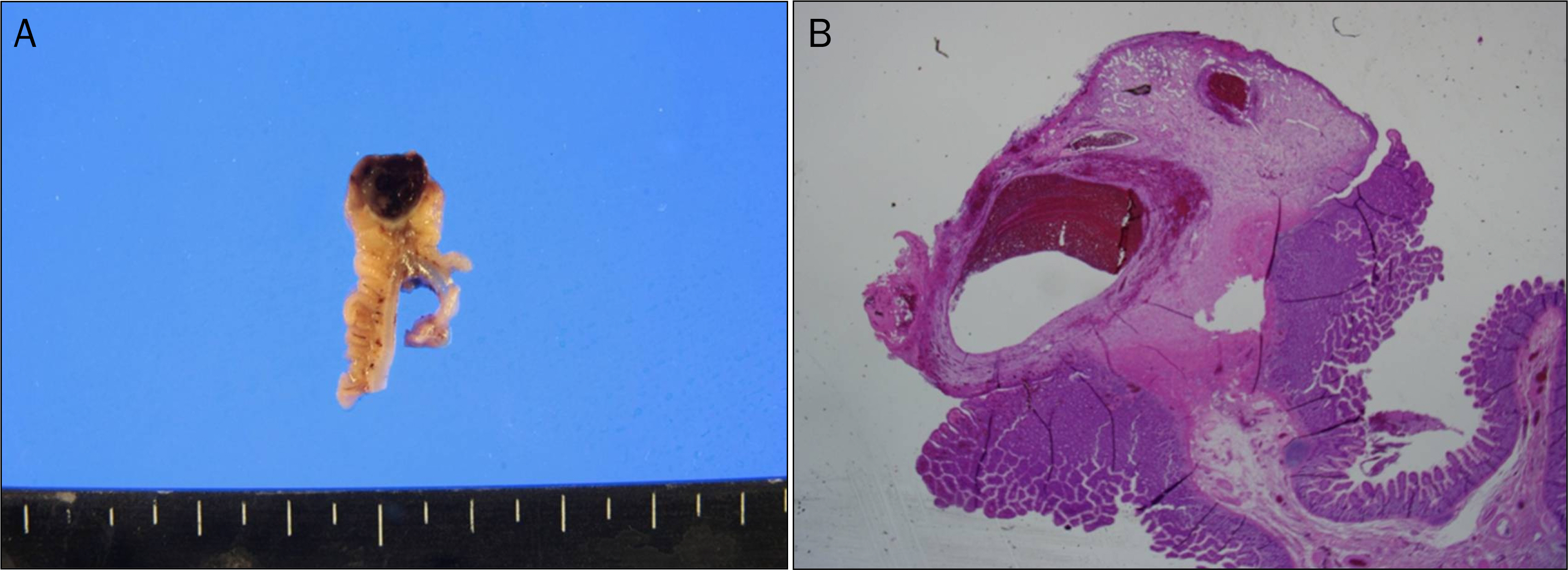
- Dieulafoy's lesions of the Jejunum are extremely rare. Therefore, localization of lesions is very difficult due to their small size and tendency of occasional bleeding. However, it is important to mention the location of the Dieulafoy's lesions to prevent excessive intestinal resections or, even worse, resection of the normal intestine. We report a case of preoperative localization of a Dieulafoy's lesion embolized by a metallic coil that allows a surgeon to accurately identify the bleeding, permitting a minimally invasive surgical treatment. A 25-year-old man presented with massive hematochezia. There was no definite bleeding focus on the upper gastrointestinal endoscopy and colonoscopy. An angiography found a persistent extravasation of the contrast media at the end of straight artery of the mid-jejunal branch, around the terminal ileum, embolized with metallic coils immediately. The combination of embolized metallic coils and intraoperative fluoroscopy allowed accurate identification and minimal laparotomy. Consequently, a highly selective and minimal resection of the jejunum containing the dieulafoy lesion was possible without any postoperative complications.
Keyword
MeSH Terms
Figure
Reference
-
References
1. Jain R, Chetty R. Dieulafoy disease of the colon. Arch Pathol Lab Med. 2009; 133:1865–1867.
Article2. Lipka S, Rabbanifard R, Kumar A, Brady P. A single-center United States experience with bleeding Dieulafoy lesions of the small bowel: diagnosis and treatment with single-balloon enteroscopy. Endosc Int Open. 2015; 3:E339–E345.
Article3. Juler GL, Labitzke HG, Lamb R, Allen R. The pathogenesis of Dieulafoy's gastric erosion. Am J Gastroenterol. 1984; 79:195–200.4. Goldenberg SP, DeLuca VA Jr, Marignani P. Endoscopic treatment of Dieulafoy's lesion of the duodenum. Am J Gastroenterol. 1990; 85:452–454.5. Dulic-Lakovic E, Dulic M, Hubner D, et al. Bleeding Dieulafoy lesions of the small bowel: a systematic study on the epidemiology and efficacy of enteroscopic treatment. Gastrointest Endosc. 2011; 74:573–580.
Article6. Navuluri R, Kang L, Patel J, Van Ha T. Acute lower gastrointestinal bleeding. Semin Intervent Radiol. 2012; 29:178–186.
Article7. Durham JD, Kumpe DA, Rothbarth LJ, Van Stiegmann G. Dieulafoy disease: arteriographic findings and treatment. Radiology. 1990; 174(3 Pt 2):937–941.
Article8. Lee YT, Walmsley RS, Leong RW, Sung JJ. Dieulafoy's lesion. Gastrointest Endosc. 2003; 58:236–243.
Article9. Sai Prasad TR, Lim KH, Lim KH, Yap TL. Bleeding jejunal Dieulafoy pseudopolyp: capsule endoscopic detection and laparoscopic- assisted resection. J Laparoendosc Adv Surg Tech A. 2007; 17:509–512.10. Athanasoulis CA, Moncure AC, Greenfield AJ, Ryan JA, Dodson TF. Intraoperative localization of small bowel bleeding sites with combined use of angiographic methods and methylene blue injection. Surgery. 1980; 87:77–84.11. Crawford ES, Roehm JO Jr, McGavran MH. Jejunoileal arteriovenous malformation: localization for resection by segmental bowel staining techniques. Ann Surg. 1980; 191:404–409.12. Fogler R, Golembe E. Methylene blue injection. An intraoperative guide in small bowel resection for arteriovenous malformation. Arch Surg. 1978; 113:194–195.
- Full Text Links
- Actions
-
Cited
- CITED
-
- Close
- Share
- Similar articles
-
- Correction: Minimal Resection of Jejuna Dieulafoy's Lesion Using an Intraoperative Fluoroscopic Localization of the Metallic Coils Used in Angiography
- Laparoscopic Resection of Bleeding Dieulafoy's Lesion in the Jejunum Following Intraoperative Localization Using Endoscopy
- Life-threatening Gastrointestinal Bleeding from a Dieulafoy’s Lesion in the Duodenum: A Case Report
- A Case of Dieulafoy Lesion of the Jejunum Presented with Massive Hemorrhage
- A Multidisciplinary approach to treat massive recurrent hematochezia from a jejunal Dieulafoy lesion: A case report


