Preoperative Radiologic Evaluation of Cholangiocarcinoma
- Affiliations
-
- 1Department of Radiology and Research Institute of Radiology, Asan Medical Center, University of Ulsan College of Medicine, Seoul, Korea. sykimrad@amc.seoul.kr
- KMID: 2383408
- DOI: http://doi.org/10.4166/kjg.2017.69.3.159
Abstract
- In patients with cholangiocarcinoma, surgical resection with curative intent is the only way to achieve cure. Since surgical resection of cholangiocarcinomas is technically demanding, determination of resectability and accurate preoperative staging are crucial. For these purposes, high quality imaging including multidetector computed tomography and magnetic resonance imaging with magnetic resonance cholangiopancreaticography, is mandatory. This article will present recent advances in imaging techniques for cholangiocarginomas, potential pitfalls in imaging evaluation, and a checklist for preoperative radiologic assessment of resectability in these patients with an emphasis on perihilar cholangiocarinoma.
Keyword
MeSH Terms
Figure
-
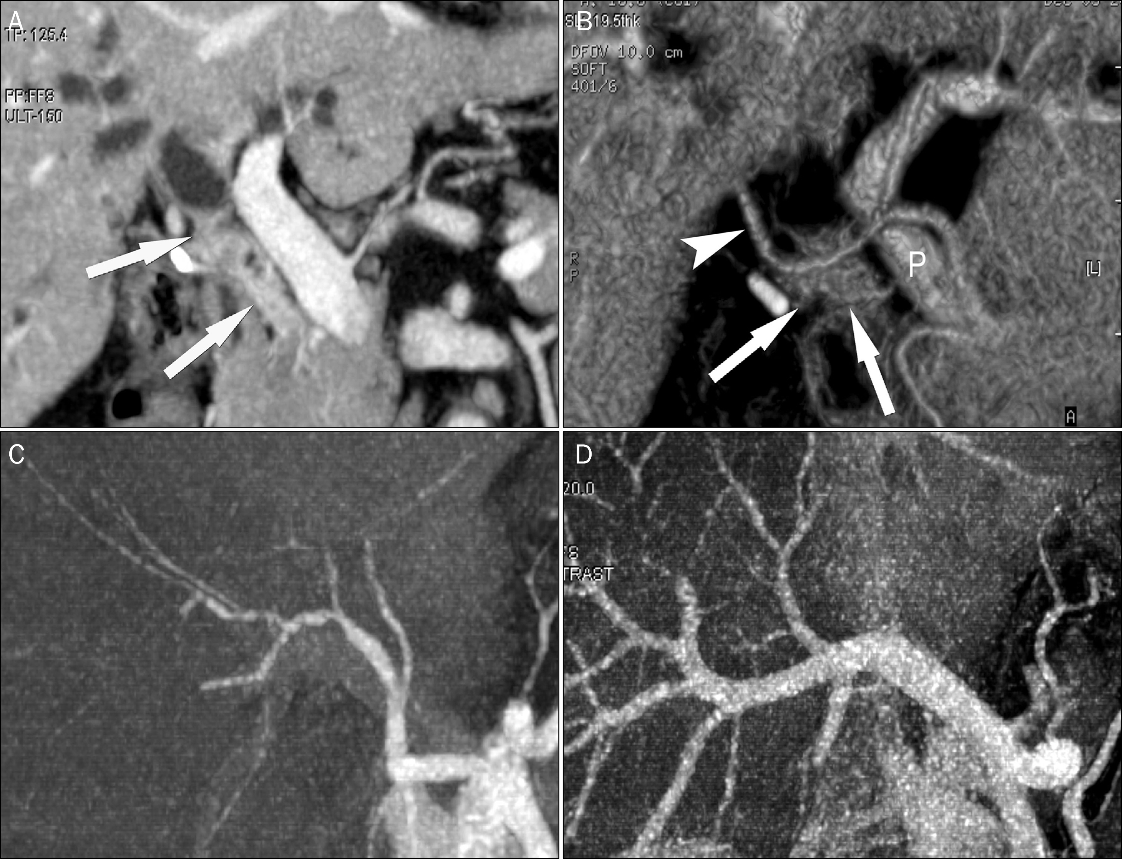
Fig. 1. MDCT images for staging CCA. (A) Coronal reformatted image demonstrates the longitudinal tumor extent (arrows) in a patient with pCCA. (B) Volume rendering image shows the relationship between the tumor (arrows) and hepatic vessels (arrowhead, hepatic artery; P, portal vein). (C, D) CT arteriography and CT venography images reconstructed with maximum intensity projection clearly depict vascular anatomy in the patient. MDCT, multidetector computed tomography; CCA, cholangiocarcinoma; pCCA, perihilar cholangiocarcinoma; CT, computed tomography.
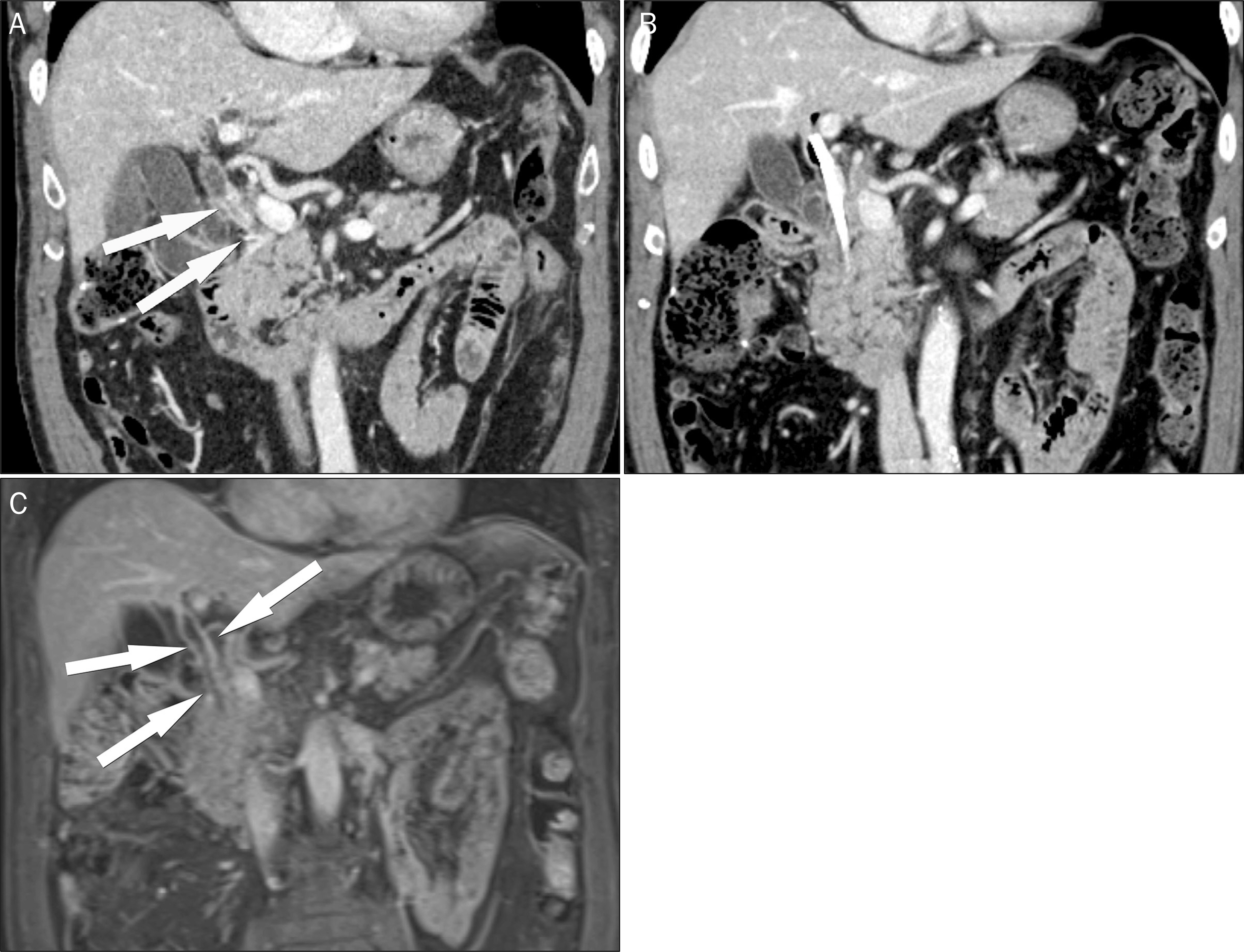
Fig. 2. Effects of biliary stent in a patient with CCA. (A) Coronal reformatted contrastenhanced CT image demonstrates the distal CCA (arrows). (B) The longitudinal tumor extent is difficult to determine in CT after biliary stent insertion. (C) On contrast enhanced coronal MR images, there is a diffuse bile duct wall thickening at the entire bile duct (arrows) due to postinverventional inflammatory changes which may cause overstaging of disease extent. CCA, cholangiocarcinoma; CT, computed tomography; MR, magnetic resonance.
Reference
-
References
1. Nakanuma Y, Curado MP, Franceschi S, et al. Intrahepatic cholangiocarcinoma. Bosman FT, Carneiro F, Hruban RH, Theise ND, editors. WHO Classification of Tumours of the Digestive System. Volume 3. 4th ed.Lyon: IARC Press;2010. p. 217–224.2. Nakanuma Y, Sato Y, Harada K, Sasaki M, Xu J, Ikeda H. Pathological classification of intrahepatic cholangiocarcinoma based on a new concept. World J Hepatol. 2010; 2:419–427.
Article3. Mar WA, Shon AM, Lu Y, et al. Imaging spectrum of cholangiocarcinoma: role in diagnosis, staging, and posttreatment evaluation. Abdom Radiol (NY). 2016; 41:553–567.
Article4. Singh MK, Facciuto ME. Current management of cholangiocarcinoma. Mt Sinai J Med. 2012; 79:232–245.
Article5. Mansour JC, Aloia TA, Crane CH, Heimbach JK, Nagino M, Vauthey JN. Hilar cholangiocarcinoma: expert consensus statement. HPB (Oxford). 2015; 17:691–699.
Article6. Sakamoto E, Nimura Y, Hayakawa N, et al. The pattern of infiltration at the proximal border of hilar bile duct carcinoma: a histologic analysis of 62 resected cases. Ann Surg. 1998; 227:405–411.7. Khan SA, Davidson BR, Goldin RD, et al. Guidelines for the diagnosis and treatment of cholangiocarcinoma: an update. Gut. 2012; 61:1657–1669.
Article8. Razumilava N, Gores GJ. Cholangiocarcinoma. Lancet. 2014; 383:2168–2179.
Article9. Joo I, Lee JM. Imaging bile duct tumors: pathologic concepts, classification, and early tumor detection. Abdom Imaging. 2013; 38:1334–1350.
Article10. Hennedige TP, Neo WT, Venkatesh SK. Imaging of malignancies of the biliary tract- an update. Cancer Imaging. 2014; 14:14.11. Ruys AT, van Beem BE, Engelbrecht MR, Bipat S, Stoker J, Van Gulik TM. Radiological staging in patients with hilar cholangiocarcinoma: a systematic review and metaanalysis. Br J Radiol. 2012; 85:1255–1262.
Article12. Seo H, Lee JM, Kim IH, et al. Evaluation of the gross type and longitudinal extent of extrahepatic cholangiocarcinomas on contrastenhanced multidetector row computed tomography. J Comput Assist Tomogr. 2009; 33:376–382.
Article13. Lee HY, Kim SH, Lee JM, et al. Preoperative assessment of resectability of hepatic hilar cholangiocarcinoma: combined CT and cholangiography with revised criteria. Radiology. 2006; 239:113–121.
Article14. Manfredi R, Barbaro B, Masselli G, Vecchioli A, Marano P. Magnetic resonance imaging of cholangiocarcinoma. Semin Liver Dis. 2004; 24:155–164.
Article15. Jhaveri KS, Hosseini-Nik H. MRI of cholangiocarcinoma. J Magn Reson Imaging. 2015; 42:1165–1179.
Article16. Park HS, Lee JM, Choi JY, et al. Preoperative evaluation of bile duct cancer: MRI combined with MR cholangiopancreatography versus MDCT with direct cholangiography. AJR Am J Roentgenol. 2008; 190:396–405.
Article17. Ryoo I, Lee JM, Chung YE, et al. Gadobutrol-enhanced, three-di-mensional, dynamic MR imaging with MR cholangiography for the preoperative evaluation of bile duct cancer. Invest Radiol. 2010; 45:217–224.
Article18. Lee MG, Park KB, Shin YM, et al. Preoperative evaluation of hilar cholangiocarcinoma with contrastenhanced three-dimensional fast imaging with steady-state precession magnetic resonance angiography: comparison with intraarterial digital subtraction angiography. World J Surg. 2003; 27:278–283.
Article19. Masselli G, Manfredi R, Vecchioli A, Gualdi G. MR imaging and MR cholangiopancreatography in the preoperative evaluation of hilar cholangiocarcinoma: correlation with surgical and pathologic findings. Eur Radiol. 2008; 18:2213–2221.
Article20. Hänninen EL, Pech M, Jonas S, et al. Magnetic resonance imaging including magnetic resonance cholangiopancreatography for tumor localization and therapy planning in malignant hilar obstructions. Acta Radiol. 2005; 46:462–470.
Article21. Fowler KJ, Brown JJ, Narra VR. Magnetic resonance imaging of focal liver lesions: approach to imaging diagnosis. Hepatology. 2011; 54:2227–2237.
Article22. Cui XY, Chen HW. Role of diffusion-weighted magnetic resonance imaging in the diagnosis of extrahepatic cholangiocarcinoma. World J Gastroenterol. 2010; 16:3196–3201.
Article23. Kang Y, Lee JM, Kim SH, Han JK, Choi BI. Intrahepatic mass-forming cholangiocarcinoma: enhancement patterns on gadoxetic acid-enhanced MR images. Radiology. 2012; 264:751–760.
Article24. Péporté AR, Sommer WH, Nikolaou K, Reiser MF, Zech CJ. Imaging features of intrahepatic cholangiocarcinoma in Gd-EOB-DTPA-enhanced MRI. Eur J Radiol. 2013; 82:e101–e106.
Article25. Bae KE, Kim SY, Lee SS, et al. Assessment of hepatic function with Gd-EOB-DTPA-enhanced hepatic MRI. Dig Dis. 2012; 30:617–622.
Article26. Huh J, Kim SY, Yeh BM, et al. Troubleshooting arterialphase MR images of gadoxetate disodium-enhanced liver. Korean J Radiol. 2015; 16:1207–1215.
Article27. Kim SY, Park SH, Wu EH, et al. Transient respiratory motion arti-fact during arterial phase MRI with gadoxetate disodium: risk factor analyses. AJR Am J Roentgenol. 2015; 204:1220–1227.
Article28. Masselli G, Gualdi G. Hilar cholangiocarcinoma: MRI/MRCP in staging and treatment planning. Abdom Imaging. 2008; 33:444–451.
Article29. Esnaola NF, Meyer JE, Karachristos A, Maranki JL, Camp ER, Denlinger CS. Evaluation and management of intrahepatic and extrahepatic cholangiocarcinoma. Cancer. 2016; 122:1349–1369.
Article30. Park HJ, Kim SH, Jang KM, Lee SJ, Park MJ, Choi D. Differentiating hepatic abscess from malignant mimickers: value of dif-fusion-weighted imaging with an emphasis on the periphery of the lesion. J Magn Reson Imaging. 2013; 38:1333–1341.
Article31. Bismuth H, Corlette MB. Intrahepatic cholangioenteric anastomosis in carcinoma of the hilus of the liver. Surg Gynecol Obstet. 1975; 140:170–178.32. Jarnagin WR, Fong Y, DeMatteo RP, et al. Staging, resectability, and outcome in 225 patients with hilar cholangiocarcinoma. Ann Surg. 2001; 234:507–517. discussion 517–509.
Article33. Deoliveira ML, Schulick RD, Nimura Y, et al. New staging system and a registry for perihilar cholangiocarcinoma. Hepatology. 2011; 53:1363–1371.
Article34. Engelbrecht MR, Katz SS, van Gulik TM, Laméris JS, van Delden OM. Imaging of perihilar cholangiocarcinoma. AJR Am J Roentgenol. 2015; 204:782–791.
Article
- Full Text Links
-
- Actions
-
Cited
- CITED
-
- Close
- Share
-
- Similar articles
-
- Hilar Cholangiocarcinoma: Recent update of radiologic assessment
- Hilar Cholangiocarcinoma
- Current Status and Recent Update of Imaging Evaluation for Peri-Hilar Cholangiocarcinoma
- The Significance of Serum and Bile Interleukin-6 Levels for Preoperative Detection of Cholangiocarcinoma in Patients with Hepatolithiasis
- Preoperative Diagnosis and Management for Hilar Cholangiocarcinoma
