A Case of Intramural Hematoma of the Esophagus Mimicking Acute Coronary Syndrome
- Affiliations
-
- 1Department of Internal Medicine, Pusan National University School of Medicine, Busan, Korea. luckyace@hanmail.net
- 2Research Institute for Convergence of Biomedical Science and Technology, Pusan National University Yangsan Hospital, Yangsan, Korea.
- 3Department of Internal Medicine, BHS Hanseo Hospital, Busan, Korea.
- 4Department of Internal Medicine, Hwa Myung Il Sin Christian Medical Center, Busan, Korea.
- 5Dr. Hong's Internal Medicine, Yangsan, Korea.
- KMID: 2383402
- DOI: http://doi.org/10.4166/kjg.2017.69.4.239
Abstract
- Intramural hematoma of the esophagus is a rare condition that can be spontaneous or secondary to trauma, toxic ingestion, or intervention. If it is the spontaneous type, it usually presents initially with epigastric pain, hematemesis or dysphagia. We present a case of intramural hematoma of the esophagus mimicking acute coronary syndrome. A 63-year-old man presented with severe acute chest pain. He has four coronary stents that were inserted five years ago, from a different hospital, and is on dual antiplatelet agents. Coronary angiography was performed immediately under the suspicion of acute coronary syndrome, and we found that there was no obvious clogging of the coronary arteries. Next, chest computed tomography was performed due to suspected aortic dissection, and the result was also negative. Four days later, endoscopy was performed and intramural hematoma covered with large ulcers was diagnosed.
Keyword
MeSH Terms
Figure
-

Fig. 1 Chest computed tomography findings. Diffuse wall thickening from the middle esophagus (A) to the cardia of the stomach (B) is observed, with eccentric thickening mainly in the medial and posterior aspect without contrast enhancement.
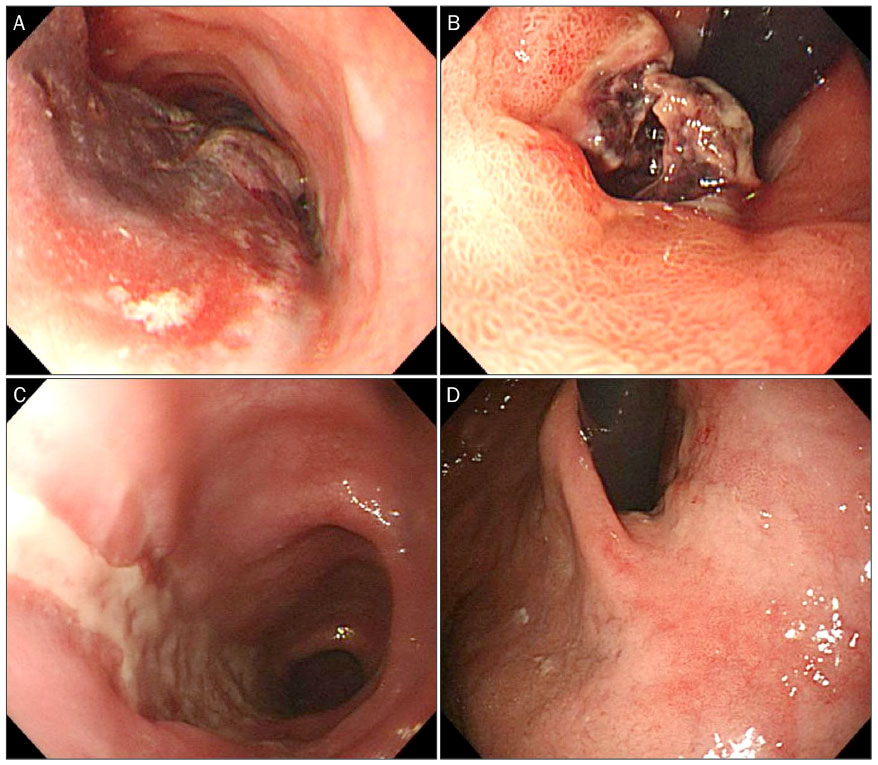
Fig. 2 Endoscopic findings. From the middle esophagus, a long red purple hematoma covered with some ulcer was observed (A), which was passed from the gastroesophageal junction to the cardia of the stomach (B). Seven days later, the hematoma has disappeared and a longitudinal ulcer with whitish plaques is observed matching the site. Mid esophagus (C) and the cardia of the stomach (D).

Fig. 3 Histopathologic finding of the esophageal wall. Inflammatory cells and normal glands are observed and there are no findings such as dysplasia (H&E, ×100).
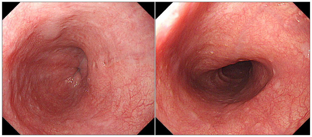
Fig. 4 Endoscopic findings of the esophagus after 8 weeks. The hematomas and ulcers have disappeared.
Reference
-
1. Steadman C, Kerlin P, Crimmins F, et al. Spontaneous intramural rupture of the oesophagus. Gut. 1990; 31:845–849.2. Koike J, Matsushima M, Teraoka H, et al. A case of submucosal hematoma of the esophagus and stomach, possibly caused by fish bone ingestion. Tokai J Exp Clin Med. 2010; 35:46–56.3. Williams B. Case report; oesophageal laceration following remote trauma. Br J Radiol. 1957; 30:666–668.4. Ko BM, Park GH, Hong SJ, et al. Spontaneous intramural hematoma of the esophagus. Korean J Gastrointest Endosc. 1998; 18:872–877.5. Lee CH, Jung HG, Kim DH. A case of spontaneous intramural hematoma of the esophagus. Korean J Gastrointest Endosc. 2010; 40:309–311.6. Cullen SN, McIntyre AS. Dissecting intramural haematoma of the oesophagus. Eur J Gastroenterol Hepatol. 2000; 12:1151–1162.7. Restrepo CS, Lemos DF, Ocazionez D, Moncada R, Gimenez CR. Intramural hematoma of the esophagus: a pictorial essay. Emerg Radiol. 2008; 15:13–22.8. Hsu CC, Changchien CS. Endoscopic and radiological features of intramural esophageal dissection. Endoscopy. 2001; 33:379–381.9. Modi P, Edwards A, Fox B, Rahamim J. Dissecting intramural haematoma of the oesophagus. Eur J Cardiothorac Surg. 2005; 27:171–173.10. Hiller N, Zagal I, Hadas-Halpern I. Spontaneous intramural hematoma of the esophagus. Am J Gastroenterol. 1999; 94:2282–2284.11. Nagai T, Torishima R, Nakashima H, et al. Spontaneous esophageal submucosal hematoma in which the course could be observed endoscopically. Intern Med. 2004; 43:461–467.12. Sen A, Lea RE. Spontaneous oesophageal haematoma: a review of the difficult diagnosis. Ann R Coll Surg Engl. 1993; 75:293–295.13. Ackert JJ, Sherman A, Lustbader IJ, McCauley DI. Spontaneous intramural hematoma of the esophagus. Am J Gastroenterol. 1989; 84:1325–1328.
- Full Text Links
-
- Actions
-
Cited
- CITED
-
- Close
- Share
-
- Similar articles
-
- A Case of Spontaneous Intramural Hematoma of the Esophagus
- Pill-induced Intramural Hematoma of the Esophagus
- A Case of Intramural Hematoma of the Esophagus Accompanied with Subcutaneous Hemorrhage
- Dissecting Intramural Hematoma of the Esophagus: A case report
- Intramural Hematoma of the Esophagus after Endoscopic Pinch Biopsy
