Squamous Cell Carcinoma Developing within Lesions of Disseminated Superficial Actinic Porokeratosis
- Affiliations
-
- 1Department of Dermatology, Eulji Hospital, College of Medicine, Eulji University, Seoul, Korea. drhams@eulji.ac.kr
Abstract
- Disseminated superficial actinic porokeratosis (DSAP) consists of multiple annular, hyperkeratotic lesions that have a bilateral distribution on sun-exposed areas, particularly the extremities. DSAPs have a wider distribution than porokeratosis of Mibelli and usually develop during the 3rd or 4th decade of life. Squamous cell carcinoma that arises in the classical type of porokeratosis of Mibelli is well-documented, but there are only a few reports of squamous cell carcinoma in DSAP. Here, we describe a 62-year-old man with DSAP who developed squamous cell carcinoma on his right forearm.
Figure
-
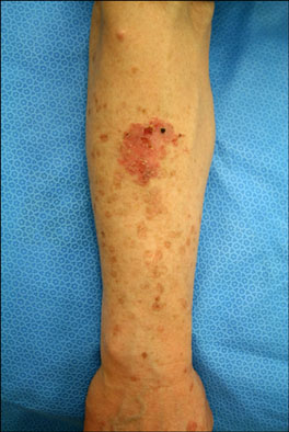
Fig. 1 An irregular, marginated, erythematous plaque and multiple, brown, atrophic macules surrounded by well-demarcated, raised ridges on the right forearm.
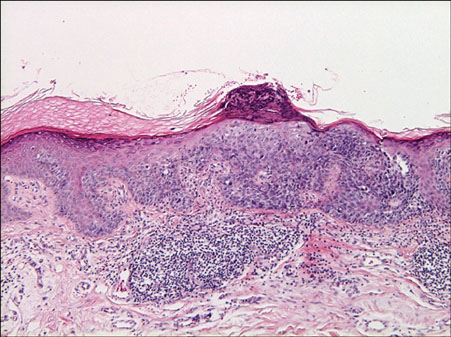
Fig. 2 Biopsy specimen obtained from the erythematous plaque on the right arm. In epidermis, acanthosis and dysregulated keratinocytes with hyperchromatic, atypical nuclei are observed. A cornoid lamella composed of a column of parakeratosis is seen in the lesion of the squamous cell carcinoma (H&E, ×100).
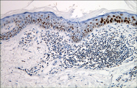
Fig. 3 Overexpression of p53 in the epidermis of a disseminated superficial actinic porokeratosis lesion. A column of parakeratosis with underlying hypogranulosis is observed. Perivascular lymphocytic infiltrations are localized beneath the cornoid lamella (p53 immunohistochemical stain, ×200).
Reference
-
1. Shumack SP, Commens CA. Disseminated superficial actinic porokeratosis: a clinical study. J Am Acad Dermatol. 1989. 20:1015–1022.
Article2. Schwarz T, Seiser A, Gschnait F. Disseminated superficial "actinic" porokeratosis. J Am Acad Dermatol. 1984. 11:724–730.
Article3. Yang HY, Nam TS, Kim YT, Kim JH. A case of squamous cell carcinoma and Bowen's disease associated with superficial disseminated porokeratosis. Ann Dermatol. 1990. 2:31–34.
Article4. James WD, Rodman OG. Squamous cell carcinoma arising in porokeratosis of mibelli. Int J Dermatol. 1986. 25:389–391.
Article5. Shrum JR, Cooper PH, Greer KE, Landes HB. Squamous cell carcinoma in disseminated superficial actinic porokeratosis. J Am Acad Dermatol. 1982. 6:58–62.
Article6. Chernosky ME, Rapini RP. Squamous cell carcinoma in lesions of disseminated superficial actinic porokeratosis: a report of two cases. Arch Dermatol. 1986. 122:853–855.
Article7. Leache A, Soto de Delás J, Vázquez Doval J, Lozano MD, Quintanilla E. Squamous cell carcinoma arising from a lesion of disseminated superficial actinic porokeratosis. Clin Exp Dermatol. 1991. 16:460–462.
Article8. Won JH, Lee MJ, Park JS, Chung H. A case of the giant and hyperkeratotic variant of porokeratosis. Korean J Dermatol. 2009. 47:101–103.9. Ninomiya Y, Urano Y, Yoshimoto K, Iwahana H, Sasaki S, Arase S, et al. p53 gene mutation analysis in porokeratosis and porokeratosis-associated squamous cell carcinoma. J Dermatol Sci. 1997. 14:173–178.
Article10. Lozinski AZ, Fisher BK, Walter JB, Fitzpatrick PJ. Metastatic squamous cell carcinoma in linear porokeratosis of Mibelli. J Am Acad Dermatol. 1987. 16:448–451.
Article11. Guss SB, Osbourn RA, Lutzner MA. Porokeratosis plantaris, palmaris, et disseminata. A third type of porokeratosis. Arch Dermatol. 1971. 104:366–373.
Article
- Full Text Links
-
- Actions
-
Cited
- CITED
-
- Close
- Share
-
- Similar articles
-
- Early Squamous Cell Carcinoma Arising from Disseminated Superficial Actinic Porokeratosis
- Various Skin Tumors Originating from Disseminated Superficial Actinic Porokeratosis
- A Case of Squamous Cell Carcinoma and Bowen's Disease Associated with Superficial Disseminated Porokeratosis
- Coexistence of Porokeratosis of Mibelli and Disseminated Superficial Actinic Porokeratosis
- A Case of Linear Porokeratosis Superimposed on Disseminated Superficial Actinic Porokeratosis
