Ann Dermatol.
2011 Nov;23(4):512-514.
Kissing Nevus of the Penis
- Affiliations
-
- 1Department of Dermatology, Chonnam National University Medical School, Gwangju, Korea. schul@chonnam.ac.kr
Abstract
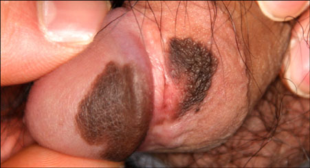
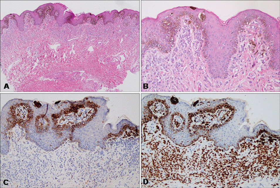
- Kissing or divided nevi are similar in shape to congenital melanocytic nevi located on an adjacent part of the body that are separated during embryogenesis. Kissing nevi of the upper and lower eyelids have been reported infrequently since the first report in 1908. Kissing nevi of the penis are very rare, with only 12 cases being reported until now, and this is the first case report in the Korean dermatological literature. A previously healthy 27-year-old man presented with asymptomatic black colored patches, which were detected 10 years ago, on the glans penis and the prepuce with growth in size. We report here a case of kissing nevus of the penis, which showed an obvious mirror-image symmetry relative to the coronal sulcus.
Figure
Reference
-
1. Fuchs A. Divided nevi of the eyelids. Urol Cutaneous Rev. 1950. 54:88–90.2. Harrison R, Okun M. Divided nevus. A clue to the intrauterine development of melanocytic nevi. Arch Dermatol. 1960. 82:235–236.3. Hamming N. Anatomy and embryology of the eyelids: a review with special reference to the development of divided nevi. Pediatr Dermatol. 1983. 1:51–58.
Article4. Desruelles F, Lacour JP, Mantoux F, Ortonne JP. Divided nevus of the penis: an unusual location. Arch Dermatol. 1998. 134:879–880.
Article5. Choi GS, Won DH, Lee SJ, Lee JH, Kim YG. Divided naevus on the penis. Br J Dermatol. 2000. 143:1126–1127.
Article6. Kono T, Nozaki M, Kikuchi Y, Erçöçen AR, Hayashi N, Chan HH, et al. Divided naevus of the penis: a hypothesis on the embryological mechanism of its development. Acta Derm Venereol. 2003. 83:155–156.
Article7. Phan PT, Francis N, Madden N, Bunker CB. Kissing naevus of the penis. Clin Exp Dermatol. 2004. 29:471–472.
Article8. Egberts F, Egberts JH, Schwarz T, Hauschild A. Kissing melanoma or kissing nevus of the penis? Urology. 2007. 69:384.e5–384.e7.
Article9. Palmer B, Hemphill M, Wootton C, Foshee JB, Frimberger D. Kissing nevus discovered at circumcision consult. J Pediatr Urol. 2010. 6:318–319.
Article10. Higashida Y, Nagano T, Oka M, Nishigori C. Divided naevus of the penis. Acta Derm Venereol. 2010. 90:319.
Article11. Zhou C, Xu H, Zang D, Du J, Zhang J. Divided nevus of the penis. Eur J Dermatol. 2010. 20:527–528.12. Sagong C, Ahn YS, Yu HJ, Kim JS. Congenital divided nevus of the eyelid. Korean J Dermatol. 2007. 45:864–866.13. Fuchs A. Ueber geteilte naevi der augenlider. Klin Monatsbl Augenheikd. 1919. 63:678–683.14. Guerra-Tapia A, Isarría MJ. Periocular vitiligo with onset around a congenital divided nevus of the eyelid. Pediatr Dermatol. 2005. 22:427–429.
Article15. Sato S, Kato H, Hidano A. Divided nevus spilus and divided form of spotted grouped pigmented nevus. J Cutan Pathol. 1979. 6:507–512.
Article16. Niizawa M, Masahashi T, Maie O, Takahashi S. A case of solitary mastocytoma suggesting a divided form of mast cell nevus. J Dermatol. 1989. 16:402–404.
Article17. Hayashi N, Soma Y. A case of epidermal nevi showing a divided form on the fingers. J Am Acad Dermatol. 1993. 29:281–282.
Article18. Demitsu T, Nagato H, Nishimaki K, Okada O, Kubota T, Yoneda K, et al. Melanoma in situ of the penis. J Am Acad Dermatol. 2000. 42:386–388.
Article19. Mandal A, Al-Nakib K, Quaba AA. Treatment of small congenital nevocellular naevi using a combination of ultrapulse carbon dioxide laser and Q-switched frequency-doubled Nd-YAG laser. Aesthetic Plast Surg. 2006. 30:606–610.
Article


